SciELO - Scientific Electronic Library Online

 
vol.57 issue2Aberrant palatal exostosisHypoplastic amelogenesis imperfecta: report of two family cases author indexsubject indexarticles search
Home Pagealphabetic serial listing  

My SciELO

Services on Demand

Journal

Article

Indicators

  • Have no cited articlesCited by SciELO

Related links

  • Have no similar articlesSimilars in SciELO

Share


Revista Cubana de Estomatología

Print version ISSN 0034-7507On-line version ISSN 1561-297X

Rev Cubana Estomatol vol.57 no.2 Ciudad de La Habana Apr.-June 2020  Epub July 13, 2020


Presentación de caso

The ice cream cone technique, a predictable treatment possibility

La técnica del cono de helado, una posibilidad de tratamiento predecible

1 Universidad de Chile, Facultad de Odontología. Departamento de Prótesis. Santiago de Chile, Chile.

2 Universidad de Ciencias Médicas de La Habana, Facultad de Ciencias Médicas “Victoria de Girón”. Departamento de Estomatología General Integral. La Habana, Cuba.

3 Práctica privada. Santiago de Chile, Chile.

4 Universidad de Chile. Facultad de Odontología. Departamento de Odontología Restauradora. Santiago de Chile, Chile.

5 Universidad Autónoma de Chile, Instituto de Ciencias Biomédicas. Santiago de Chile, Chile.

ABSTRACT

Introduction:

The volumetric preservation of bone and soft tissue after a tooth extraction has special relevance in the esthetic zone when it will be rehabilitated by a dental implant.

Objective:

Describe the prosthodontics treatment in a socket with advanced buccal bone resorption, with a flapless technique for guided bone regeneration and with a dental implant and implant-supported single fixed prosthesis.

Case presentation:

A case is presented of a male 62-year-old partially dentate patient. Radiographic examination showed the presence of advanced buccal bone resorption in relation to the maxillary left lateral incisor. It was a result of the root displacement secondary to root fracture. In a first surgical phase the lateral incisor was extracted using an atraumatic periotome technique. Particulate cortical bone allograft was compacted into the site to fill the space that was previously occupied by the root of the tooth. Temporary restoration was performed using the extracted natural tooth, which was adhesively bonded to the adjacent teeth. Four months after grafting the extraction site showed an adequate height and width of the bone. In a second surgical phase, an implant was placed. Six months after implant placement, osseointegration was clinically confirmed and a provisional crown was screwed on the implant performed. The final restoration with a zirconium dioxide abutment and a full ceramic crown was obtained and cemented.

Conclusions:

Regeneration of the buccal plate was possible through the use of particulate cortical bone allograft and a resorbable collagen membrane adapted to the bone defect and placed in a position to recreate the buccal plate. This allowed the installation of an implant 4 months later, the procedure allowing esthetic and functional results using a single fixed prosthesis.

Keywords: dental implant; surgical flap; guided tissue regeneration; tooth socket

RESUMEN

Introducción:

La preservación volumétrica de los huesos y tejidos blandos después de una extracción dental tiene especial relevancia en la zona estética cuando será rehabilitada por un implante dental.

Objetivo:

Describir el tratamiento de prótesis en una cavidad con reabsorción ósea bucal avanzada, con una técnica sin colgajo para la regeneración ósea guiada y con un implante dental y una prótesis fija única con soporte de implante.

Presentación del caso:

Paciente masculino de 62 años, edente parcial. El examen radiográfico mostró la presencia de reabsorción ósea bucal avanzada en relación con el incisivo lateral superior izquierdo. Fue el resultado del desplazamiento de la raíz secundario a la fractura de esta. En una primera fase quirúrgica, el incisivo lateral se extrajo utilizando una técnica de periotoma atraumático. El aloinjerto de hueso cortical particulado se compactó en el sitio para llenar el espacio que anteriormente ocupaba la raíz del diente. La restauración temporal se realizó utilizando el diente natural extraído, que se unió adhesivamente a los dientes adyacentes. Cuatro meses después del injerto, el sitio de extracción mostró una altura y anchura adecuadas del hueso. En una segunda fase quirúrgica, se colocó un implante. Seis meses después de la colocación del implante, se confirmó clínicamente la osteointegración y se realizó una corona provisional atornillada al implante. La restauración final con un pilar de dióxido de circonio y una corona de cerámica completa se obtuvo y se cementó.

Conclusiones:

La regeneración de la placa bucal fue posible mediante el uso de aloinjerto de hueso cortical particulado y una membrana de colágeno reabsorbible adaptada al defecto óseo y colocada en una posición para recrear la placa bucal. Esto permitió la instalación de un implante 4 meses después. El procedimiento permitió la estética y los resultados funcionales utilizando una única prótesis fija.

Palabras clave: implante dental; colgajo quirúrgico; regeneración tisular guiada; alveolo dental

Introduction

The volumetric preservation of bone and soft tissue after a tooth extraction has special relevance in the aesthetic zone when it will be rehabilitated by a dental implant. In type 2 defects, this extraction socket is defined as having the presence of facial soft tissue with a partial or complete dehiscence of the buccal bone plate,1,2 a variety of alveolar preservation techniques have been proposed to preserved or recreated the original dimensions of the ridge when the tooth was present. In these techniques, bone graft materials have been used with and without barrier membranes, however, it has not yet been defined with has better predictability. Currently, the flapless ice cream cone technique described by Elian et al.,1 has shown very good results; in some studies with this technique 1,3,4,5,6 they found very low losses in bone thickness after alveolar preservation.

Due to this, the objective of this paper was to describe the prosthodontics treatment in a socket with advanced buccal bone resorption, with a flapless technique for guided bone regeneration (GBR) and with a dental implant and implant-supported single fixed prosthesis.

Case presentation

62-year-old male patient, with stands for American Society of Anesthesiologists Classification 2 and arterial hypertension medicated with Enalapril 20 mg/day, partially dentate. The radiographic exam showed the presence of advanced buccal bone resorption in relation to the maxillary left lateral incisor (Tooth 2.2). It was a result of the root displacement secondary to root fracture (Fig. 1, A). The patient’s main complaint was aesthetic.
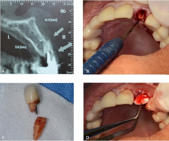
Fig. 1-  A: Cone Beam Computer Tomography (CBCT) images tooth 2.2. B: Tooth extracted using an atraumatic periotome technique. C: Root and crown extracted. D: A resorbable collagen membrane was adapted to the bone defect and placed in position to recreate the buccal plate. 

In a first surgical phase the lateral incisor was extracted using an atraumatic periotome technique (Fig. 1, B and C). A resorbable collagen membrane (BioMend Extend, Zimmer Biomet Dental, Palm Beach Gardens, FL) (15 mm x 20 mm) was adapted to the bone defect and placed in position to recreate the buccal plate (Fig. 1, D). Particulate cortical bone allograft (Puros Cortical Allograft Particulate, Zimmer Biomet Dental) (0,5 cc) was compacted into the site to fill the space that was previously occupied by the root of the tooth. The membrane sealed the space wrapping to palatal gingiva and it was retained in position with three horizontal mattress sutures (Fig. 2, A and B). Temporary restoration was performed using the extracted natural tooth, which was adhesively bonded to the adjacent teeth (Fig. 2, C and D).
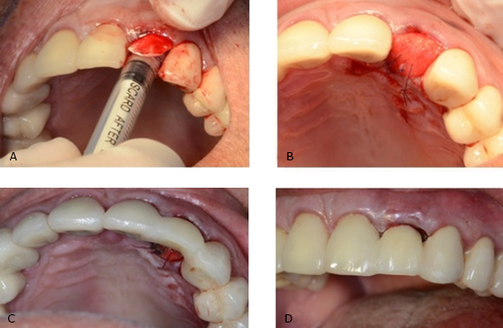
Fig. 2-  A and B: The membrane sealed the space wrapping to palatal gingiva and it was retained in position with three horizontal mattress sutures. C and D: Temporary restoration was performed using the extracted natural tooth, which was adhesively bonded to the adjacent teeth. 

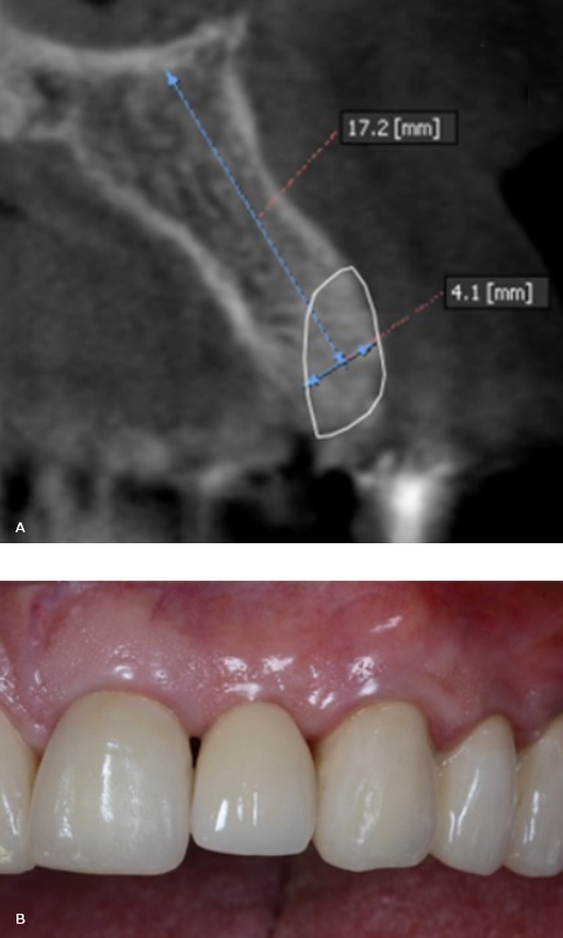
Four months after grafting the extraction site in preparation for dental implant placement, conventional radiology and CBCT showed an adequate height and width of the bone (Fig. 3, A).

In a second surgical phase, one implant was placed (Screw-Vent, Zimmer Biomet Dental) (3.3 x 16 mm) with an average surgical torque of 35 Ncm. Six months after the implant placement was radiographically controlled, osseointegration was clinically confirmed and a provisional crown screwed on the implant was performed. After several stages, the final restoration with a zirconium dioxide abutment and a full ceramic crown was obtained and cemented (Fig. 3, B).

Fig. 3-  A: Four months after grafting the extraction site in preparation for dental implant placement, conventional radiology and CBCT showed an adequate height and width of the bone. B: Final restoration with a zirconium dioxide abutment and a full ceramic crown. 

Discussion

The ice cream cone technique for alveolar preservation used in this clinical situation, showed to be adequate to rebuilt and maintenance the remain buccal plate in type 2 defects. This prevents the collapse of the buccal tissue plate creating a new buccal bone plate and then avoiding the aesthetic problem that causes. In fact , researches about the buccolingual dimensional changes of the extraction socket using the “Ice Cream Cone” technique conclude that this technique is an easy approach that results in better horizontal regeneration of the buccal bone plate.6

This technique avoids making flaps with the possibility of soft tissue retraction, in addition to being a relatively easy practice, which allows reducing the operative times for the clinician and patients.1,5

In addition, bone regeneration with this technique allowed an adequate surgical torque and thus be able to perform an immediate temporary crown.

Although, research on this technique is still limited, the studies that have been conducted so far have shown satisfactory results1,6 in relation to other procedures described like “Cytocone Procedure”.3 This is a modification of the original Ice cream cone technique”. A nonresorbable d-PTFE barrier is placed in and over a socket tipe II that has received a bone graft without a buccal flap being raised. The author of the paper said that for this technique Its major benefit compared to the original technique is the use of a nonresorbable barrier (ie, d-PTFE).7 This eliminates the possibility of premature barrier resorption by salivary enzymes, which can cause loss of bone graft material, interrupt the process of GBR, and result in a deformed ridge vertically or horizontally.3,7

GBR is based on the principle that a barrier membrane is used for space maintenance over a defect, promoting the ingrowth of osteogenic cells and preventing migration of undesired cells from the overlying soft tissues into the wound. Protection of a blood clot in the defect, exclusion of gingival connective tissue, and provision of a secluded space into which osteo- genic cells from the bone can mi- grate are essential for a successful outcome.6

Because the Cytocone technique uses a nonresorbable barrier, the clinician is able to dictate how long the barrier remains in place. Usually it is retained for 5 to 6 weeks to ensure predictable GBR. This interval allows enough time for osteoid formation and connective tissue coverage of the socket opening. After barrier removal, the epithelium migrates across the connective tissue within 2 weeks. Epithelium travels approximately 0.5 mm to 1 mm per day.3

As previously discussed, if the ice cream cone technique is performed with a resorbable barrier, the barrier may degrade prematurely, which can result in incomplete GBR. In contrast, the Cytocone procedure assists bone regeneration without compromising esthetics; and, furthermore, patients have minimal discomfort.3

Clinicians have several choices with respect to treatment planning a case if an implant is going to be inserted to replace a hopeless tooth that is adjacent to buccal bony dehiscence. After an extraction, a GBR procedure (bone graft plus a barrier) can be accomplished with a traditional nonresorbable barrier (e-PTFE) in conjunction with primary closure, or a d-PTFE or resorbable barrier can be employed without primary closure like as was showed in original ”Ice cream cone technique”.1 As discussed, the ice cream cone technique or the Cytocone procedure can be used to regenerate bone prior to placing an implant, or implantation can be performed in conjunction with a GBR procedure.1,2,3 All of these procedures have advantages and limitations.

Other modifications from the original technique have been proposed to try to optimize the results.2,8

Conclusions

The regeneration of the buccal plate was possible through the use of particulate cortical bone allograft and a resorbable collagen membrane adapted to the bone defect and placed in a position to recreate the buccal plate. This allowed the installation of an implant (3.3 mm x 16 mm) 4 months later the procedure. This technique allowed esthetics and functional results using a single fixed prosthesis.

Bibliographic references

1. Elian N, Cho SC, Froum S, Smith RB, Tarnow DP. A simplified socket classification and repair technique. Pract Proced Aesthet Dent [Internet]. 2007 [acceso 07/07/2019];19(2):[aprox. 7 p.]. Disponible en: https://www.ncbi.nlm.nih.gov/pubmed/17491484Links ]

2. Sarnachiaro GO, Chu SJ, Sarnachiaro E, Gotta SL, Tarnow DP. Immediate Implant Placement into Extraction Sockets with Labial Plate Dehiscence Defects: A Clinical Case Series. Clin Implants Dent Related Res [Internet]. 2016 [acceso 07/07/2019];18(4): [aprox. 9 p.]. Disponible en: https://onlinelibrary.wiley.com/doi/abs/10.1111/cid.12347Links ]

3. Changi K, Greenstein G. Cytocone Procedure: Conservative Repair of a Buccal Plate Dehiscence in Preparation for Implant Placement. Compendium of Continuing Education in Dentistry (Jamesburg, NJ: 1995) [Internet]. 2018 [acceso 07/07/2019]; 39(5):[aprox. 6 p.]. Disponible en: https://europepmc.org/abstract/med/29714495Links ]

4. Ten Heggeler J, Slot D, Van der Weijden G. Effect of socket preservation therapies following tooth extraction in non-molar regions in humans: a systematic review. Clin Oral Implants Res [Internet]. 2011 [acceso 07/07/2019];22(8):[aprox. 10 p.]. Disponible en: https://onlinelibrary.wiley.com/doi/abs/10.1111/j.1600-0501.2010.02064.xLinks ]

5. Al-Juboori MJ. Conservative Socket Regeneration with Buccal Wall Defect Using Guided Tissue. Open Dent J [Internet]. 2016 [acceso 07/07/2019];10:[aprox. 7 p.]. Disponible en: https://www.ncbi.nlm.nih.gov/pmc/articles/PMC5093866/pdf/TODENTJ-10-561.pdfLinks ]

6. Tan-Chu JH, Tuminelli FJ, Kurtz KS, Tarnow DP. Analysis of buccolingual dimensional changes of the extraction socket using the "ice cream cone" flapless grafting technique. Int J Period Restorative Dent [Internet]. 2014 [acceso 07/07/2019];34(3):[aprox. 5 p.]. Disponible en: https://doi.org/10.11607/prd.1605Links ]

7. Soldatos NK, Stylianou P, Koidou VP, Angelov N, Yukna R, Romanos GE. Limitations and options using resorbable versus nonresorbable membranes for successful guided bone regeneration. Quintessence international (Berlin, Germany : 1985) [Internet]. 2017 [acceso 07/07/2019];48(2):[aprox. 17 p.]. Disponible en: http://qi.quintessenz.de/index.php?doc=abstract&abstractID=37133/Links ]

8. Basualdo J, Ivankovic M, Kuzmicic J, Fernández E. Atraumatic Extraction and immediate implant placement into infected site with the "ice cream cone" technique and L-PRF: A Case Report. Rev Clin Periodoncia Implantol Rehabil Oral [Internet]. 2018 [acceso 07/07/2019];11(1):[aprox. 4 p.]. Disponible en: https://scielo.conicyt.cl/scielo.php?pid=S0719-01072018000100043&script=sci_arttext&tlng=enLinks ]

Received: July 05, 2019; Accepted: October 19, 2019

*Corresponding author: chaple@infomed.sld.cu

The authors not declared conflict of interests.

This case presentation was not received neither financial support by any institution.

Creative Commons License This is an open-access article distributed under the terms of the Creative Commons Attribution License