


My SciELO
 Custom services
Services on Demand
Article
 English (pdf)
 Article in xml format
 Article references
 Send this article by e-mail
Indicators
-
 Cited by SciELO
Related links
-
 Similars in
SciELO
Similars in
SciELO
Share

 Permalink
Revista de Salud Animal
Print version ISSN 0253-570X
Rev Salud Anim. vol.36 no.3 La Habana Sept.-Dec. 2014
ORIGINAL ARTICLE
Serological prevalence and risk factors of Borrelia burgdorferi in water buffaloes (Bubalus bubalis, Linnaeus, 1758) on Marajó Island, northern region of Brazil
Prevalencia serológica y factores de riesgo de Borrelia burgdorferi en búfalos de agua (Bubalus bubalis, Linnaeus, 1758) en la isla Marajó, región norte de Brasil
Jenevaldo Barbosa da SilvaI*, Bruna de A. BaêtaII, Cinthia T.A. LopesIII, Bruna Sampaio Martins Land ManierII, Gustavo Nunes Santana de CastroII, Priscilla Nunes dos SantosII, Adivaldo Henrique da FonsecaII, José Diomedes BarbosaIII
IFaculdade de Ciências Agrárias e Veterinárias - UNESP, Rod. Carlos Tonanni, km 05, 14870-000, Jaboticabal, São Paulo, Brazil. E-mail: jenevaldo@hotmail.com.
IIUniversidade Federal Rural do Rio de Janeiro - UFRRJ, BR465, Km 07, 23890-000, Seropédica, Rio de Janeiro, Brazil. E-mail: adivaldofonseca@yahoo.com.
IIIUniversidade Federal do Pará, Centro Agropecuário, Departamento de Ciência Animal. Rua Maximino Porpino da Silva, 1000, Centro Castanhal, PA, Brazil. 68748-080. E-mail: diomedes@ufpa.br.
ABSTRACT
Sera samples were collected from 330 water buffaloes on Marajó Island, state of Pará, Brazil, to assess the presence of antibodies against Borrelia burgdorferi by indirect Enzyme Linked Immunosorbent Assay. Approximately 45% of the animals had antibodies against B. burgdoferi. The prevalence of seropositive buffaloes, 72% (85/118), was statistically higher in the city of Soure than in the other municipalities tested. Murrah breed animals were significantly more seropositive (Prevalence Ratio (PR) = 1.84, p = 0.000) than those of the Mediterranean breed. Among the animals diagnosed positive for tuberculosis, 33% (4/12), were also seropositive for B. burgdoferi. Animals positive for tuberculosis had a significantly lower level of B. burgdorferi seropositivity (PR = 1.36, p = 0.0017) than negative animals. The study showed that there was an agent immunologically close to B. burgdorferi circulating in water buffalo on Marajó Island, state of Pará, Brazil. These results demonstrate a possible role of the water buffalo in the epidemiological chain of this agent in Brazil, and are a warning of the danger this disease could pose to public health.
Key words: spirochetes, Borrelia sp., ELISA, buffaloes, risk factors.
RESUMEN
Para determinar la presencia de anticuerpos contra Borrelia burgdorferi por iELISA, se colectaron 330 muestras de suero de búfalos de agua en la Isla Marajó, estado de Pará, Brasil. Aproximadamente el 45% de los animales tuvieron anticuerpos contra B. burgdoferi. La prevalencia de búfalos seropositivos, 72% (85/118), fue estadísticamente más alta en la ciudad de Soure que en el resto de las municipalidades estudiadas. Los animales de raza Murrah fueron significativamente más positivos (radios de prevalencia (RP) = 1.84, p = 0.000) que los de la raza Mediterranean. Entre los animales diagnosticados positivos para tuberculosis, 33% (4/12) también fueron seropositivos para B. burgdoferi. Los animales positivos para tuberculosis tuvieron un nivel significativamente menor de seropositividad para B. burgdorferi (PR = 1.36, p = 0.0017) que los animales negativos. El estudio mostró que existe un agente inmunológicamente relacionado con B. burgdorferi circulando en búfalos de agua en la isla Marajó, estado de Pará, Brasil. Estos resultados demuestran el posible papel del búfalo de agua en la cadena epidemiológica de este agente en Brasil y que constituye una advertencia sobre el daño que esto puede ocasionar a la salud pública.
Palabras clave: espiroquetas, Borrelia sp., ELISA, búfalos, factores de riesgo.
INTRODUCTION
In 1890, the water buffalo arrived in Brazil at Marajó Island, Pará state; today they are found throughout the country and constitute the largest herd in the West (1), with one third of the national herd still remaining on the island of Marajó (2).
The genus Borrelia (Swellengrebel, 1907) consists of Gram negative, microaerophilic, mobile bacteria, classified in the order Spirochaetales and family Spirochaetaceae. Lyme borreliosis, caused by spirochetes belonging to the Borrelia burgdorferi sensu lato complex, is an anthropozoonosis referred to as Baggio-Yoshinari syndrome in Brazil (3). Ticks act as vectors, becoming infected upon ingestion of blood from an infected vertebrate host, and showing transovariarial and transtadial transmission (4, 5, 6). Wild animals, including deer, rodents and birds, represent important reservoirs of the spirochete (7, 8).
Serological analysis by Enzyme Linked Immunosorbent Assay (ELISA) has a wide application in epidemiological studies of Lyme borreliosis because a large number of samples can be concomitantly analized at low cost; however, it has low specificity and cross reactions may occur (9, 10).
In Brazil, previous studies have confirmed the presence of antibodies against B. burgdorferi in several animal species such as cattle (11, 12), horses (13), and dogs (14, 15). The first report of Borrelia sp. in water buffaloes was from the state of Pará, when Scofield et al. (16) observed morphometric spirochetes in a buffalo with clinical suspicion of Enzootic Bovine Leukosis. In 2012, Corrêa et al. (17) found 83.9% of buffaloes seropositive for B. burgdorferi; however, the possible risk factors for infection are still unknown. Therefore, the objective of this study was to use indirect ELISA to evaluate the presence of antibodies against B. burgdorferi in water buffaloes on Marajó Island, state of Pará, and their association with potential risk factors.
MATERIALS AND METHODS
The study was conducted in the municipalities of Soure, Muaná, Salvaterra, Thailand Ponta de Pedra, and Santa Cruz Arari during the year 2012. All the municipalities belong toMarajó Island, Pará, located at latitude 00º 43'00 «south and longitude 48º 31'24 «west, with an altitude of 10 meters.
Blood samples were collected by coccygeal vein puncture from 330 randomly selected buffaloes aged between two and three years. Of these animals, 156 were of Mediterranean and 174 of Murrah breed. Among all animals studied, 123 were pregnant. Twelve buffaloes tested positive for tuberculosis, and 15 were positive for brucellosis.
The enzyme-linked immunosorbent assay was based on the description of Silva et al. (12). The antigen was prepared from B. burgdorferi sensu stricto G 39/40 of North American origin and adjusted to 5 µg/ml protein to coat plates by overnight incubation at 4°C, after which the plates were blocked for 90 minutes at 37°C. Test samples were diluted 1:400 in PBS-Tween 20 with 5% milk powder, and incubated in plates at 37°C in a humid chamber for 90 minutes. After this, 100µl of the alkaline phosphatase anti-bovine IgG (Sigma Chemical Co.), diluted to 1:30000 according to manufacturer's recommendations, was added to each well. The plates were incubated in the same conditions as before. The substrate p-nitrophenyl phosphate (PNPP, Sigma Chemical Co.) was added at 100µl/well and the plates were incubated at room temperature for 50 minutes, after which the absorbance was read at 405 nm in a micro-ELISA reader (Labsystems iEMS Reader MF).
Cut-off values were calculated based on 30 non-Borrelia-infected water buffalo sera by the receiver operating characteristic (ROC) analysis with MedCalc statistical software (version 11.4; http://www.medcalc.be) (18). Thirty serum samples obtained from foals before colostrum suckling were used as negative controls. A positive reference group consisting of 10 buffalo serum samples tested positive for B. burgdorferi (titres of 1280 by IFAT) were used as positive controls in the serological assays.
The chi-square test was used to determine significant differences in the percentages of samples tested, at a significance level of 5%. The association between seropositivity to B. burgdorferi and risk factors of locality, breed, physiological state (pregnancy) and positivity to tuberculosis and brucellosis was performed using the chi-square (÷2). The analysis was performed by the statistical software R Foundation for Statistical Computing, version 2.12.2 (2011).
RESULTS
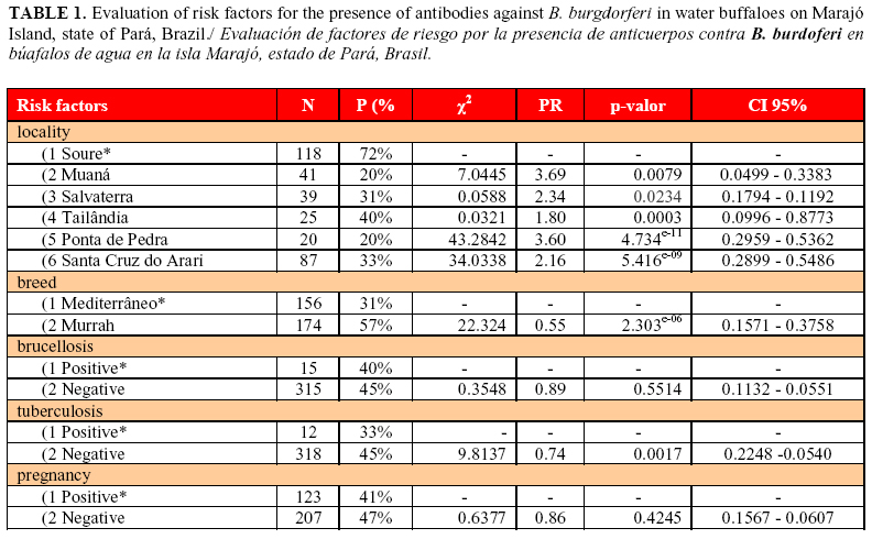
Among the 330 buffalo serum samples analyzed, 148 animals had antibodies against B. burgdoferi, representing a prevalence of 44.8%. The number of seropositive animals varied significantly (p<0.05) depending on the location, breed and positivity for tuberculosis (Table 1).
The prevalence of seropositive buffalo found in the city of Soure (72%) was statistically higher than in the other municipalities studied. Murrah animals were significantly more seropositive (PR = 1.84, p = 0.000) than the Mediterranean breed animals. Among animals positive for tuberculosis, 33% (4/12) were also seropositive for B. burgdoferi, a significantly lower level of seropositivity (PR = 1.36, p = 0.0017) than that seen in tuberculosis-negative animals (Table 1).
DISCUSSION
The number of seropositive animals observed in this study indicates the presence of a spirochete immunologically similar to B. burgdorferi in buffaloes in the region studied, as previously suggested by Scofield et al. (16) and Corrêa et al. (17). The frequency of seropositive buffaloes in this study was lower than the frequency observed by Corrêa et al. (17) in working with Murrah buffalo from the mainland and Marajó Island, in the state of Pará, Brazil. This study showed that Murrah buffaloes were more likely to have antibodies against B. burgdorferi than the Mediterranean breed, explaining the lower rate of positive animals seen here, when both breeds were studied, compared with Corrêa et al. (17). In a study on the same region, Guedes-Junior et al. (11) found a similar seroprevalence (54.9%) of B. burgdorferi in cattle.
In a study of cattle, Kocan et al. (19) found a limitation in the use of the European breed Bos taurus, it does not have antibodies against tick-borne diseases. In contrast, Bos indicus, of Indian origin, has a greater degree of resistance, because of the immunity it acquired, for its greater contact with agents. The same idea can be applied to the Mediterranean and Murrah breeds of buffalo, since the Mediterranean breed of European origin, as with Bos taurus, showed lower seropositivity, whereas Murrah animals (Indian origin) had higher levels of seropositivity than B. burgdorferi.
The drop in immunity caused by tuberculosis (20) impairs the immune response against other agents - this could explain the association observed between animals positive for tuberculosis and seronegative against B. burgdorferi.
CONCLUSIONS
This study demonstrates the existence of an immunologically similar agent to B. burgdorferi circulating in water buffaloes on Marajó Island, state of Pará, Brazil. The lower prevalence of antibodies against B. bugdorferi in buffaloes positive for tuberculosis may indicate the importance of this debilitating disease in impairing the immune response not only to borreliosis, but possibly to other agents as well. Future studies on buffaloes should consider the importance of mixed infections by different agents.
Differential breed susceptibility is already known in cattle, but there have not been similar studies on buffaloes. Although we observed that Murrah buffaloes were more susceptible than the Mediterranean buffaloes, more detailed studies should be conducted to evaluate the immunological basis of this finding.
These results demonstrate the possible role of the buffalo in the epidemiological chain of this disease in Brazil, and serve as a warning of the danger that this disease poses to public health.
CONFLICT OF INTEREST STATEMENT
None of the authors of this work has a financial or personal relationship with other people or organizations that could inappropriately influence or bias the content of the paper.
ACKNOWLEDGEMENTS
We are grateful to the Coordination for the Improvement of Higher Level of Education Personnel (CAPES) and National Counsel of Technological and Scientific Development (CNPq) for financial support.
REFERENCES
1. Bernardes O. A bubalinocultura no Brasil: situação e importância econômica. Rev Bras Reprod Anim. 2007;31(2):293-298.
2. IBGE (Instituto Brasileiro de Geografia e Estatística). Disponível em: <http://www.ibge.gov.br> Acesso em: 01 jun 2013.
3. Mantovani E, Costa IP, Gauditano G, Bonold VLN, Higuchi ML, Yoshinari NH. Description of Lyme disease-like syndrome in Brazil. Is it a new tick borne disease or Lyme disease variation? Braz J Med Biol Res. 2007;40(1):443-456.
4. Smith RD, Brener J, Osorno M, Ristic M. Pathobiology of Borrelia theileri in the tropical cattle tick, Boophilus microplus. J Invertebr Pathol. 1978;32(1):182-190.
5. Hoogstraal H. Argasid and nuttalliellid ticks as parasites and vectors. Adv Parasitol. 1985;24(1):135-238.
6. Lane RS, Burgdorfer W. Transovarial and transstadial passage of Borrelia burgdorferi in the western black-legged tick, Ixodes pacificus Acari: Ixodidae. Am J Med Hyg. 1987;37(1):188-192.
7. Steere AC. Lyme disease. N Engl J Med. 1989;31(1):586-597.
8. Bosler EM. Tick vectors and hosts. In: Coyle PK. Eds. Lyme Disease. Mosby Year Book, Boston. 1993:18-26.
9. Rogers AB, Smith RD, Kakoma I. Serologic cross-reactivity of antibodies against Borrelia theileri, Borrelia burgdorferi and Borrelia coriaceae in cattle. Am J Vet Res. 1999;60(1):694-697.
10.Magnarelli LA, Bushmich SL, Sherman BA, Fikrig E. A comparison of serologic tests for the detection of serum antibodies to whole-cell and recombinant Borrelia burgdorferi antigens in cattle. Can Vet J. 2004;45(1):667-674.
11.Guedes-Junior DS, Araújo FR, Silva FJM, Rangel CP, Barbosa Neto JD, Fonseca AH. Frequency of antibodies to Babesia bigemina, B. bovis, Anaplasma marginale, Trypanosoma vivax and Borrelia burgdorferi in cattle from the northeastern region of the state of Pará, Brazil. Rev Bras Parasitol Vet. 2008;17(1):105-109.
12.Silva JB, Baêta BA, Ribeiro CCDU, Teixeira RC, Fonseca AH. Detection of antibodies against Borrelia burgdorferi in periparturient cows and calves during the first year old by indirect enzyme-linked immunosorbent assay iELISA. GJSFR. 2012;13(1):15-19.
13.Galo KR, Fonseca AH, Madureira RC, Barbosa Neto JD. Frequência de anticorpos homólogos anti- Borrelia burgdorferi em equinos na mesorregião metropolitana de Belém, Estado do Pará. Pesq Vet Bras. 2009;29(1):229-232.
14.Alves AL, Madureira RC, Silva RA, Corrêa FN, Botteon RCCM. Frequência de anticorpos contra Borrelia burgdorferi em cães na região metropolitana do Rio de Janeiro. Pesq Vet Bras. 2004;24(1):203-206.
15.Cordeiro MD, Meireles GS, Silva JB, Souza MMS, Fonseca AH. Soroprevalência para Borrelia spp. em cães no município de Seropédica, estado do Rio de Janeiro. Rev Bras Med Vet. 2012;34(1):251-256.
16.Scofield A, Marques CC, Barbosa JD, Fonseca AH. Ocorrência de Borrelia sp. em búfalos Bubalus bubalis no município de Castanhal, estado do Pará. In: Congresso Brasileiro de Buiatria, Anais. Buzios. 2005. pp.130.
17.Corrêa FN, Teixeira RC, Oliveira CMC, Barbosa JD, Fonseca AH. Detection of anti-Borrelia burgdorferi antibodies in buffaloes Bubalus bubalis in the state of Pará, Brazil. Rev Bras Parasitol Vet. 2012;21(1):338-341.
18.Terkawi MA, Huyen NX, Shinuo C, Inpankaew T, Maklon K, Aboulaila M, et al. Molecular and serological prevalence of Babesia bovis and Babesia bigemina in water buffaloes in the northeast region of Thailand. Vet Parasitol. 2011;78(1):201-207.
19.Kocan KM, Fuente J, Guglielmone AA, Melendez RD. Antigens and alternatives for control of Anaplasma marginale infection in cattle. Clin Microbiol Rev. 2003;16(1):698-712.
20.Rodrigues CA, Medeiros E, Mello GC, Favaro MR. Controle da Tuberculose Bovina. Rev Cient Eletr Med Vet. 2008;11:1-5.
Recibido: 30-1-2014.
Aceptado: 17-7-2014.
*Correspondencia: Jenevaldo Barbosa da Silva. E-mail: jenevaldo@hotmail.com.
