


My SciELO
 Custom services
Services on Demand
Journal

Article
 English (pdf)
 Article in xml format
 Article references
 Send this article by e-mail
Indicators
-
 Cited by SciELO
Related links
-
 Similars in
SciELO
Similars in
SciELO
Share

 Permalink
Biotecnología Aplicada
On-line version ISSN 1027-2852
Biotecnol Apl vol.26 no.4 La Habana Oct.-Dec. 2009
REVIEW
Micro-magnetic and molecular magnetic resonance imaging in modern biotechnology and pharmacy
Imágenes moleculares y microimágenes de resonancia magnética en la biotecnología y la farmacéutica modernas
Carlos Cabal, Evelio González, Yulenia Torne, Angel Rojas
Unidad de Resonancia Magnética, Centro de Neurociencias de Cuba, CIREN. Ave. 25 No. 15202 esq. 158, Playa, Ciudad de La Habana, Cuba
ABSTRACT
Magnetic Resonance Imaging is an essential diagnostic technique for the non-invasive study of biological entities with high contrast as well as high spatial and temporal resolution. This work describes the current state of the art in Micro-Magnetic Resonance Imaging and Molecular Magnetic Resonance Imaging. These new technologies are transforming the modus operandi of the current research and applications being performed and developed in the biotechnological and medical-pharmaceutical industries; establishing metabolic and genotypic relationships to phenotypical traits and validating the design of active molecules, their structure-function relationship and their interaction with the intended target. The use of endogenous and exogenous contrast mechanisms is also described. Therefore, underlining the importance, a special emphasis is being placed on these methods capable of detecting, quantifying and visualizing dynamic molecular and cellular phenomena in tissues and organs at spatial resolutions of the order of dozens of microns and temporal resolutions of milliseconds. Examples of the application of these methods in the study of pathologies like cancer and brain stroke are shown. The development of recent advances in hardware, devices and contrast mechanisms is referred to increase productivity, specificity, reliability and impact in experimental work. In the near future, the study of these techniques will be an indispensable tool for the design, development and validation of new pharmaceuticals.
Keywords: Molecular images, magnetic resonance, biotechnology, pharmaceutical industry
RESUMEN
Las imágenes de resonancia magnética son esenciales para estudiar de modo no invasivo la naturaleza biológica, con elevado contraste y alta resolución temporal y espacial. Se describe el estado actual del arte de las microimágenes y de las imágenes moleculares de resonancia magnética, nuevas ideas transformadoras de las investigaciones y aplicaciones en la biotecnología y la industria medico-farmacéutica que constituyen una herra-mienta efectiva para el establecimiento de la relación metabólica y genotípica con la expresión fenotípica y para comprobar diseños de moléculas activas así como su relación estructura-función e interacción con blancos terapéuticos. Se expone el uso de mecanismos contrastantes endógenos y exógenos. Se destaca la importancia de estos métodos que detectan, cuantifican y visualizan fenómenos dinámicos moleculares y celulares en tejidos y órganos, con resoluciones espaciales de hasta decenas de micras y temporales, de milisegundos. Se detalla su utilización para el estudio de afecciones como la isquemia cerebral y el cáncer. Se hace referencia a los avances obtenidos en los equipos, dispositivos, metodologías y mecanismos de contraste para elevar la productividad, especificidad, fiabilidad e impacto de las investigaciones. En breve tiempo, los estudios por imágenes serán ineludibles para el diseño, desarrollo y validación de nuevas drogas.
Palabras clave: Imágenes moleculares, resonancia magnética, biotecnología, industria farmacéutica
INTRODUCTION
Many of the promises of Magnetic Resonance Imaging (MRI) for the non-invasive study of live tissues at high temporal resolutions with unmatched spatial resolution and contrast have become a reality in current biochemical and preclinical research, as well as in clinical and industrial settings. These potentialities, however, are far from exhausted (1-5).
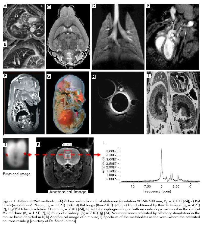
The present work aims at summarizing the current state of the art in two emerging MRI methods which are transforming a number of research conceptions and opening up new ideas for practical applications in biotechnology and medical-pharmaceutical industry: Magnetic Resonance microimages (μMR) and molecular images (miMR). Although the definitions of these concepts are not completely settled yet, the following constitutes good approximations: μMR is the technique to produce images from live biological structures using magnets in the order of a few tens of microns, with high contrast and short acquisition times, allowing the study of anatomical details and physiological processes of cells, tissues, and organs without discernible alterations (Figure 1). On the other hand, miMR, are in vivo, non-invasive magnetic resonance techniques for the visual representation, structural characterization and quantification of biological processes at cellular and molecular levels (6). Although μMR and miMR could be in fact are being applied for research at the molecular, cellular and tissue levels in botany and agricultural biotechnology, such applications will not be discussed here (7).
In addition, μMR and miMR play a decisive contribution in genomic evaluation and link metabolomics, proteomics and phenotypic traits together unraveling the bases of different diseases (2, 4, 7, 8). They constitute an effective complement for the evaluation of molecularly designed active compounds, studying their structure-function relationship and their interaction with the intended therapeutic targets (2-14).
In toxicological studies, μMR and miMR are playing a rising significant role by complementing and accelerating these assays and increasing the speed and reliability of the results of the pharmacokinetic evaluation of new therapeutic molecules at the molecule, cell, tissue and organism levels (2-14). These methods guarantee higher accuracy, reducing the required amounts of reagents and laboratory animals, changing the labor-intensive nature of histological work, and shortening sampling intervals during model evaluation and quantification of experimental parameters; consequently lead to an increased speed and lowered costs. The accuracy of longitudinal and transversal studies is notably favored as well by limiting the need to euthanize experimentation animals and comparing the model to itself, (14, 15).
Although all of them provide useful and unique information to a higher or lower extent, making a comparison of the spatial resolution, image acquisition time, maximum assay depth, equipment costs and possibilities of different imagenological modalities reveals that only MRI can provide molecular, cellular, anatomical and physiological information with unmatched spatial resolution and a comparable temporal resolution. Another advantage of MRI, not often explicitly outlined in the literature, is the direct application to the clinical practices of procedures, technology and software originally developed for lab animal and vice versa, taking into account that the experimental bases of MR data acquisition in both situations are very similar. The main disadvantage is MRI equipment cost, which remains more expensive unlike the alternative methods (12).
The number of scientific publications dealing with μMR and miMR has exponentially increased in the last 10 years (16), and the number of reports evaluating favorably the position of regulatory entities worldwide on these technologies is also being increased in frequency (13-15). It is predicted that the requirements for the study, validation and registration of new pharmaceuticals will include the use of images and data from image processing in a few years. Therefore, the introduction, use and development of these image acquisition technologies in all fields of biotechnology and biomedicine is becoming necessary to remain competitive at the international level.
MAGNETIC RESONANCE METHODS
The phenomenon of magnetic resonance (MR) is based on the processes of polarization (alignment) and excitation of nuclei from atoms with a magnetic moment that are commonly found in living beings (1H, 13C, 14N, 23Na, 31P…). In order to align these nuclei, the studied body is introduced into a homogeneous magnetic field of relatively high intensity (B0). Excitation is achieved by subjecting the experimental sample to radiofrequency electromagnetic wave pulses (RF) that perturb the aligned nuclei from their equilibrium position. Once the RF pulse stops, the return of the nuclei to the equilibrium position defined by the magnetic field can be characterized by two parameters, called relaxation times T1 and T2. In turn, T1 and T2 depend on the biophysical and biochemical properties (density, molecular structures and movement, biological barriers, temperature, pH…) of all elements of the sample, from single molecules to whole organs. The intensity of the MR signal (intensity of each pixel for MRI) is a function of the density of magnetic nuclei (ρ) and T1 y T2, and therein lays the information that can be provided by MRI (1). Hydrogen (1H) is the most commonly used nucleus in MRI due to its abundance (guaranteeing the most intense signal) and forms part of H2O and most molecular structures. Thus, it is the signal registered in MRI originates from water molecules in its different states. Changes in concentration, mobility, associations and interactions of H2O are involved in all normal or pathological and physiological processes, consequently registered in MR and MRI signals (17).
The acquisition of MRI images involves the synchronous application of RF pulses and carefully selected magnetic field gradients (G). There are many image acquisition modalities, some stated below, arising from specific combinations of RF pulses, G and times to trigger the previous mentioned above relaxation processes (1). Different MRI processes offer different degrees of detail and precision in the exa-mination of the specific molecular, cellular or tissular phenomena under study, and MRI machines both for clinical or animal experimentation often store libraries of pulse sequences for these purposes, providing the researcher a wide array of choices in spatial reso-lution, signal/noise ratio and image acquisition time. On the other hand, there are atlases registering bi-dimensional (2D) and three-dimensional (3D) images of human and rodent organs (18).
Weighted imaging in ρ, T1 and T2
Classical MRI can be biased towards water density (ρ) , T1 or T2, and these techniques are known as: ρ-, T1- or T2-weighted MRI. All pathological processes are associated with variations in the content and mobility of water, resulting in changes to T1 and T2 during MRI (17). During the first years of biomedical application of T1-, T2- and ρ-weighted MRI proved their potential for the detection of cerebral lesions in animal models of ischemia, tumors, post-traumatic wounds and multiple sclerosis (2, 4, 5). A growth in hydrogen density (H2O), T1 and T2 in pathological tissues is attributed to an increase of interstitial water associated to the development of vasogenic edema. As it has been demonstrated, in rat and gerbil brains T1 and T2 correlate well with water content, and changes in this parameter are more intense reflected in T2.Moreover, changes in T2 are more pronounced in tumoral edemas in experimentation animals. The higher sensitivity of T2-weighted images has lead to their wide use in experimental and clinical settings (4, 5). During early stages of acute ischemia in animals models, the changes in T1 and T2 can be attributed to a decreased tissue oxygenation (T2-weighted MRI) and the interruption of blood flow (T1-weighted MRI) as already demonstrated.
Diffusion-weighted imaging
One of the most powerful and promising methods of MRI is the use of diffusion-weighted imaging (DWI), where each pixel of the image reflects the intensity and direction of movement of water molecules in each microzone of the object under examination.H2O, subjected to molecular movement either as a free molecule or as part of the hydration spheres of other molecules and ions, changes the motility of its molecular partners depending on the nature of the molecular, cellular and physiological processes involved in.
The diffusion at each pixel (with volumes ranging from a few dozen picoliters to some microliters) depends on bio-structural factors such as obstacles or barriers (e.g. cell membranes, blood-brain barrier, ma-cromolecules, etc). The diffusion coefficient (D) of H2O varies significantly when it is measured either along or across such barrier, which is reflected in pi-xel intensity in MR images. Indeed, using the mean apparent D per pixel (ADC, Apparent Diffusion Co-efficient) it is possible to determine the density and orientation of biological barriers for each micro-region by DWI, or more specifically, ADC images (17).
Measuring diffusion requires the application of an additional magnetic field gradient, whose orientation allows exploring the location and alignment of the barriers mentioned above. In order to obtain ADC information and define barrier orientation at least two consecutive images have to be acquired, which in turn requires restraining severely the movement of the experimental subject to avoid compromising data quality due to involuntary motion (1, 17).
DWI has opened up a wide array of experimental choices, given its increased sensitivity to cell variations and tissue anomalies as well as the possibility of detecting affected zones with higher resolution and at earlier stages than with T1- or T2-weighted imaging. ADC values, for instance, suffer a significant decrease of 30 to 40% in DWI just a few minutes after cerebral ischemia (4, 5), and it is possible to detect decreases in ADC during the evolution of cytotoxic edemas at tributed to the movement of water from the extracellular zone towards intracellular compartments with a high density of diffusion barriers.
A fast and intense decrease in ADC takes place during anoxic depolarization. The advance of the depolarization of the ischemic tissue into the border zone of the ischemia is joined by a transitory inflammation detectable by fast ADC imaging. These temporary perturbations of the tissue contribute to expand the lesion. DWI has enabled measuring lesion volume and monitoring its evolution (2, 4, 5, 19).
DWI has being used for monitoring the efficacy of anticancer therapy with cytotoxic drugs by following tumor response, using for instance cells of the 9L glioma. Thus, therapy can decrease cell density in the tumor and increase intercellular spaces, as well as water mobility and diffusion (as evidenced by their higher sensitivity during imaging) (4, 5, 20).
The barrier sensitivity of DWI has allowed the development of maps of the orientation, size and integrity of the fibrous structures of white matter that can be used to infer and follow neuronal connectivity in three dimensions: the tractography or architecture of neural tracks. The results in this field have been nothing short of spectacular, and the technique holds a vast potential for the study of the central nervous system (21).
In vivo spectroscopy
Magnetic Resonance Spectroscopy (MRS) was the first of the MR methods that provided molecular information in vivo. This technique can be performed in vitro, in vivo and with perfused organs or tissues. MRS has slowly become an essential component of the toolbox of current pharmaceutical research, especially in the in vivo metabolomics of ischemia and cerebral infarction (5), degenerative joint disorders, cardiovascular disease (3), cancer, and in respiratory and skin disorders. Figure 1L shows MRS data for the metabolites of a neuronal region (Figure 1J) belonging to a mouse brain under olfactory stimulation.
MRS allows measuring the concentration and distribution of administered drugs, as well as their interaction with the target. It also offers information about metabolic processes taking place in tissues and organs (2, 3, 5, 22-24); for instance, the identities and concentrations of many important metabolites as measured by 1H MRS easily found in the literature. Many of thesemetabolites play an essential role in the previously sta-ted phenomena involved with the development of different disorders, as exemplified by the concentrations of N-acetyl aspartate (NAA) and lactates (Lac) during hypoxic ischemia (NAA considered a neuronal integrity marker, whereas lactate is a marker of anaerobic metabolism that decreases immediately after ischemic damage and NAA increases). Furthermore, MRS is u-sed for the measurement of brain temperature (2, 5, 22, 23), and its possibilities are easily expanded with the use of multi-nuclear spectroscopic methods that are commonly used for researchers, examining not only 1H nuclei, but also 31P, 23Na, 19F and 133Cs nuclei which are present in biological systems or in the supplied drugs.
The main disadvantage of in vivo spectroscopy compared to MR-based imaging methods is related to the concentration of 1H registered by the technique. Imaging methods deal with water concentrations up to 100 moles, whereas the concentration of most metabolites is in the order of a few dozen millimoles. This implies that the spatial resolution of spectroscopic methods is much lower when voxel dimensions are in the order of centimeters (2, 5).
The use of perfusion-weighted imaging (PWI) should be included in the MRI arsenal, which using internal contrast or external contrasting agents, provides quantitative information about essential parameters of in vivo research such as: tissue perfusion, medium transit time and volume fraction occupied by blood in the tissue, among others. These parameters and the obtained from MR angiography have being vital for the elucidation of cerebrovascular processes and disorders as well as for the study of drug efficacy in different tissues (2, 5, 19).
Functional magnetic resonance
Functional magnetic resonance imaging (fMRI) is based on the detection of changes in blood magnetism as its oxygenation status changes (BOLD/ Blood Oxygen Level Dependent). Oxyhemoglobin (diamagnetic) becomes deoxyhemoglobin (paramagnetic) due to cellular metabolism, which is translated into a shortened T2 and, therefore, lower pixel intensities where oxygenation levels change more intensely.
fMRI is not only being used in clinical practice for some years now (2, 5, 13, 25); but also found a place in experimental research with neuronal models. For instance, the acquisition of two consecutive images, firstly with the subject at rest (basal state) and secondly during stimulation (auditory, visual, olfactory, chemical, etc.) can be used to pinpoint the brain areas activated by stimuli through the use of image subtraction (Figure 1J). Further, the relationship between the effects of administered drugs and brain activity has also inferred from BOLD signals in the presence or absence of stimuli (13, 25). The sensitivity of fMRI to oxygenation status has been widely applied to the analysis of cerebrovascular processes in animals during ischemia or hemorrhage, likewise to the evaluation of recovery through tissue re-oxygenation after reperfusion (13, 25).
MOLECULAR IMAGES AND CONTRAST MECHANISMS
Non-invasive in vivo miMR is defined as the visualrepresentation, characterization (structural and func-tional) and quantification of biological processes at cellular and molecular levels. This and other definitions have a point in common which the image shows a biological process at the molecular or cellular level quantifiable and occurs in vivo and in a non-invasive manner.
The overlap of advances in miMR with molecular and cellular biology technologies have generated an expanding discipline with notable impacts in the early detection of a number of diseases, treatment individualization and the development of new pharmaceutical and biotechnological products (2, 4, 6, 8, 10-14). One of the distinguishing advantages of the application of MRI to biological systems is the excellent contrast achieved for soft tissues (1-8), given its dependence on density and, essentially, the mobility of water flowing in or out of different structures. This level of contrast far exceeds the obtained in lab animals or humans by any other technique (12).
The mobility of water molecules changes by several orders of magnitude when they are associated to a macromolecule; therefore, water mobility changes depending on the physiological states of cells, tissues and organs (12, 17). New experiments, procedures and techniques developed in the last two decades have expanded the array of MRI contrast methods that can be used to underline the differences between otherwise similar biological structures, their temporal evolution and their dependence with physiopathological processes better.
Contrast during MRI can be managed and increased endogenously (intrinsic) or by agents which are exogenous (extrinsic) to the biological system.
Endogenous contrast mechanisms
Endogenous contrast mechanisms are based on the induction of differences in magnetization state or in the use of pre-existing natural differences produced by biomolecular processes, using pulse sequences specifically designed to capture into an image processes such as diffusion, blood perfusion into tissues or the flow of different biological fluids.
The most common known MR method taking into account endogenous contrast is fMRI which can visualize the delivery of oxygen to the nervous tissue by monitoring the change of oxy- to deoxyhemoglobin. Other methods, such as perfusion weighing (PWI) or flow techniques, make similar use of endogenous mechanisms to achieve image contrast (2, 5, 19).
Recent advances in genetic engineering have provided new ways to study and increase contrast and consequently dissect enzymatic process; perform cell labeling, and other tasks (26). MRI contrast can be genetically controlled through the directed expression of a contrasting protein or RNA without the need for synthesis of an exogenous agent (26). For instance, a gene for the intracellular expression of a metalloprotein forming a complex with endogenous metal ions can fulfill this role. As a matter of fact, there are significant numbers of applications using endogenous Fen+ (the most abundant paramagnetic ion in biological systems) for this purpose (26). One of the simplest procedures is the modulation of protein-associated Fen+ contents. Ectopic ferritin (Ft), for example, has been used as an artificial contrast agent (CA) in mice brain by changing its expression levels in vivo (26) through the introduction of Ft genes using viral transfection, obtaining higher amounts of Fen+ than in control cells without detectable toxicity. Fen+ from the heme group of myoglobin has also been overexpressed in transgenic animals to be used as a CA in muscular tissue, although in this case the results have not been satis-factory probably due to insufficient expression levels or a rather small efficiency of relaxation (26).
Other totally endogenous procedures for increasing contrast are based on the use of enzyme systems as those taking into consideration the genetic control of the accumulation of melanin which is a biopolymeric pigment that collects paramagnetic ions and exerts a large influence on MRI contrast, and is synthesized by tyrosine hydroxylase from tyrosine during the initial steps of the biosynthetic pathway for dopaquinone. Better contrast has been achieved by overexpressing tyrosine hydroxylase in human cell cultures, thus increasing the affinity for metal ions (26).
Exogenous contrast mechanisms
The use of exogenous contrast agents has constituted an important part of the established clinical practices for more than 20 years (4, 27, 28). They are usually paramagnetic substances that change the magnetic properties of neighboring tissues, cells or molecules, and are mostly delivered intravenously. Exogenous CAs have been successfully employed in MR studies of brain, liver, digestive system, lymphatic system, mammary glands, kidneys and cardiovascular system. Gadolinium-Diethylene triamine pentaacetic acid and Gadolinium-1,4,7,10 tetraazacyclododecane 1,4,7,0 triacetic acid (Gd(DTPA) and Gd (DOTA), respectively) constitute the most commonly used exogenous CA in clinical practices as biomolecular research and applications (4, 27, 28). Essentially, the use of exogenous CA is the need to achieve a high enough concentration in the interest area while keeping its lower concentration as possible in unrelated areas. Gdcommonly used exogenous CA in clinical practices as biomolecular research and applications (4, 27, 28). Essentially, the use of exogenous CA is the need to achieve a high enough concentration in the interest area while keeping its lower concentration as possible in unrelated areas. Gdcommonly used exogenous CA in clinical practices as biomolecular research and applications (4, 27, 28). Essentially, the use of exogenous CA is the need to achieve a high enough concentration in the interest area while keeping its lower concentration as possible in unrelated areas. Gd
The magnetization state of the CA and its nearby can change depending on the physiological processes taking place in biological structures. This property has been used for the study of enzyme activity, genetic expression, protein association, the activation of cellular pH and Ca2+ and partial O2 and CO2 pressure at the cell level (8, 10, 28). The CA is designed so that all coordination bonds of the Gd3+ ion must be occupied e.g. with sugars not modifying miMR contrast but blocking access to H2O. However, these sugars are endowed with functional groups that can interact with a specific enzyme such as β-galactosidase. This interaction produces a “slit” into the Gd3+ envelope that allows the introduction of water into the coordination sphere, immediately changing the intensity of the pixels corresponding to the microregion where the enzyme reaction takes place. This concept has been successfully used for the in vivo monitoring of gene expression in Xenopus laevis (28, 29). Intracellular Ca2+ is important for signal transduction; therefore, exploited for the design of another intracellular CA; activated in the presence of micromolar Ca2+ concentrations. Similar mechanisms have also been used to study the presence of Zn2+ ions (28). There are CAs that allow the examination of extracellular pH in healthy and cancer tissue, as stated by miMR (28).
A very promising approach of research is the use of magnetization transfer from hyperpolarized agents, particularly noble gases, which increase signal/noise ratio and contrast; allowing the study of the respiratory system (Figure 1D) and other structures with high anatomical-functional complexities (30).
An AC of particular interest for the study of the nervous system is the paramagnetic manganese ion, Mn2+, which –like Gd– produces significant reductions in T1 and T2 for the protons of water molecules and its nearby (31, 32). Manganese is a heavy metal, well known for being a cofactor of enzymes such as: superoxide dismutase (31-33), pyruvate carboxylase (34) and glutamine synthetase (31). Mn2+ can enter into excited nervous cells using some of the transport mechanisms for Ca2+, and its intracellular behavior has been well studied (31). Unlike Gd-based CAs, it can cross the blood-brain barrier and penetrates into neurons.
Three of the most relevant applications of Mn2+ as CA in MRI of the Central Nervous System are (31):
· The study of brain architecture: dissecting the connectivity between different brain areas. Recent research in rodents (35-39), primates and insects have proved the usefulness of MR with Mn2+ contrast for unraveling the cytoarchitecture of the brain.
· The identification of neuronal tracts: as a consequence of its defined movement inside the neurons, making possible its follow up to define the architecture of neuronal channels in the limited regions of interest where Mn2+ was locally injected. The first neu-ral tract studies were performed in mice, where they allowed the identification of olfactory and visual channels (31). One of the most spectacular applica-tions of miMR with Mn2+ contrast has been studying the changes in dimensions and connectivity of different regions of the brain in association with cognitive processes and brain plasticity (40, 41).
· The determination of the neuronal excitation regions: by penetrating into excited cells and allowing the use of miMR for determining the active regions independently from hemodynamic parameters, with better spatial accuracy than classic fMRI.
However, one disadvantage of Mn2+, is its high toxicity. Whether or not it becomes a mainstay CA for miMR will depend on the development of techniques for achieving an optimal distribution in the interest region at the proper dosage, with minimal residence times and proper experimental design.
Another exogenous contrast medium: magnetic nanoparticles
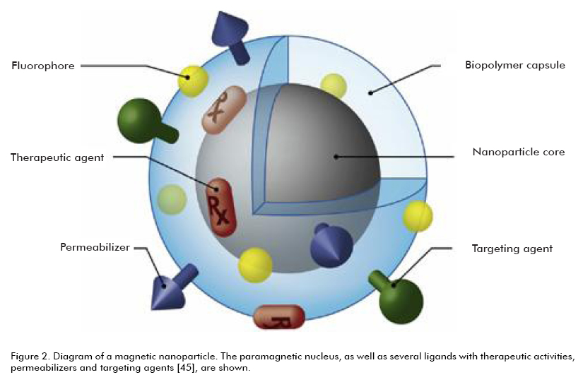
Magnetic nanoparticles (mnp), also known as superparamagnetic particles, will transform the current diagnostic and therapeutic landscape for miMR (27, 42). Their biocompatibility, bioavailability and biodegradability have extended their use to the detection, diagnosis and treatment of a diverse array of disorders such as: cerebrovascular disease, cancer and cardiovascular disease (evaluation of ischemic risk, myocardial lesions, atherosclerosis, detection and characterization of atheroma plaques and other vascular dysfunctions) (3, 5, 23, 27, 43). Mnp are classified according to their dimensions: larger or smaller than 50 nm are defined as: SPIO and USPIO, respectively (27). Their potential are given by the possibility to obtain them in a variety of shapes and chemical compositions, using a modular design to assume functions simultaneously as a label, homing and tracking device, and drug carrier and dispenser. Nanoparticles not only increase the efficacy of a number of pharmaceuticals, but have been shown to increase contrast notably during miMR, easing their microlocalization through time (27, 42).
Adequate biocompatibility, biodistribution, stability and pharmacokinetics (and hence, their versatility) are achieved by coating the mnp with specific biomolecules and biopolymers. This coating can be modified to contain therapeutic agents, homing molecules, permeabilizers and fluorescent dyes (27, 42) (Figure 2).
One of the advantages of mnp, as drug carriers, is their high surface to volume ratio, translated in a high number of therapeutic molecules per particle (Figure 2). Target ligands such as: proteins, peptides, aptamers and small molecules have been analyzed as an additive to this coating to increase the local mnp concentration at the target site. Mnp are currently essential for expanding the possibilities of miMR for real time monitoring of drug design and the use of combined therapy strategies.
The biggest limitation of most chemical therapeutic agents is their relative non-specificity and potential adverse effects on healthy tissues. Nevertheless, the high penetrability of magnetic fields into biological tissues and the development and maturity achieved by technologies for the remote management of magnetic fields in desired configurations have enabled researchers and clinicians alike to precisely steer the mnp into the desired therapeutic target, as well as to visualize and differentiate the physiological states of cells and tissues (44, 45).
The physicochemical properties of a carrier mnp (dimensions, morphology, electrical charge, chemical surface), the intensities and geometry of magnetic fields, the depth at which the diseased tissue is located, the speed of blood flow and the local level of vascularization are important parameters for this new drug delivery and release technology (45, 46).
Mnp have also contributed to raise the efficiency of antiseptic procedures and gene therapy through their control via miMR. The use of mnp carrying oligodeoxynucleotides or gene vectors overcomes many of the current limitations of gene therapy, which are merely associated with the release of the therapeutic agents (47, 48). Mnp have also been used as carriers for siRNA, one of the most promising therapeutic platforms to emerge during the last years (49, 50). Whole cells can also be labeled, tracked and guided using miMR by ‘doping’. In fact, USPIO have been successfully incorporated into stem cell populations.
Recent developments have allowed obtaining multi-functional super-paramagnetic mnp doped with fluorescent dyes that can be used for their simultaneous detection at the cellular level by MRI and optical fluorescence microscopy. In this case, MRI is used during surgical planning, and the surgical procedure is assisted and guided with both techniques to define and remove tumoral areas (51) (Figure 3). Mnp-assisted miMR have been also used to examine cellular migration and traffic, to detect apoptosis and to quantify different enzyme activities (27, 42).
STUDIES OF CANCER AND CEREBRAL ISCHEMIA
miMR have allowed monitoring with high sensitivity and specificity the behavior of important molecular targets and cancer cells during the primary and early stages of carcinogenesis (9, 10, 14, 20, 51-53).
The use of mnp for improving the detection, diagnosis and therapy of solid tumors has been extensively studied, associating it to a cytotoxic agent to turn them into a magnetic target carrier, or MTC. Thus, the intravenous injection of a colloidal suspension of the mnp, joined to the use of an external magnetic field gradient can be used to guide effectively the therapeutic agent to the target area. The accuracy of MRI to detect malignant lymphadenopathy has been improved through the use of ferumoxtran-10 mnp (54-56). A normal lymph node absorbs the USPIO agent, therefore is hypointense during MRI, being easily differentiated from a metastasized node. This phenomenon has been used for breast, lung, brain and cervical cancers, etc. On the other hand, USPIO particles have allowed the effective identification of lymphoma nodules with 5 to 10 mm diameters (57). Currently, it is possible to distinguish tumoral lesions of 2 to 3 mm in liver during clinical diagnosis with MRI associated to the use of SPIO mnp (58). Many other works are referred to angiogenesis, quantification of tumo-ral volume and delimitation of its boundaries at the cell level (Figure 3). Multiple mnp formulations are being tested in clinical trials; one of the most advanced along the clinical pipeline is Combidex, being at the last trial stages for the lympho detection of metastasis in prostate cancer (42).
Monoclonal antibodies (mAb) were the first target ligands associated to mnp, and remain the most commonly used and monitored by miMR. Their large dimensions and inherent immunogenicity constitute the biggest limitations for their use; and their conjugation to mnp limits even more their diffusion through biological barriers.
Mnp have been evaluated as carriers for a wide array of therapeutic agents. Traditional drugs as etoposide, doxorubicin and methotrexate have been encapsulated into mnp for the potential treatment of diseases ranging from rheumatoid arthritis to malignant prostate and breast tumors (59-62). Their possibilities as carriers for therapeutic proteins and peptides have also been analyzed (62), as in the case of Herceptin™ into mnp-doped liposomes which effectiveness has been demonstrated at inhibiting the proliferation of tumor cells from breast cancer, specifically releasing Herceptin™ into NIH3T.7 cells expressing HER2/neu in vivo. Another example is the use of Chlorotoxin-doped mnp (Chlorotoxin is a peptide with a high affinity to a variety of tumors which can inhibit the invasion of tumors such as gliomas (63)) increasing the therapeutic effect comparing the free peptide (64).
A great amount of publications has been published in the last years trying to analyze the involved mechanisms of post-ischemic cerebral damage or focus on developing strategies to minimize its repercussions (19, 22, 24, 65-76). Many studies in rats have evidenced the effectiveness of MRI for the detection and follow-up of lesions arising from cerebrovascular accidents (72-78). For example, it is possible to clearly pinpoint the location of the infarction in Wistar rats where ischemia has been induced by medial cerebral artery occlusion (MCAO), measuring the volume of the affected area and its evolution at 3, 7 and 24 weeks after the accident.
The most recent research on animal models of cerebral ischemia has brought about data on the role of apoptosis and cellular necrosis in this disease, monitoring the phagocytic activity and microglia in the infarcted tissue and homeostasis at the blood-brain barrier.
The evolution of the infarction after different pharmacological treatments and the inflammatory response following a cerebrovascular accident have been suc-cessfully followed and measured by MRI. A recent study (19) induced infarction in 4 different groups of animals, including a control group and three different groups receiving the intravenous administration of thrombolytic agents such as microplasmin and tissular plasminogen activator (tPA), respectively, at two different dosages. The study used different MRI methods (perfusion, diffusion, T2) to monitor after 1 and 24 hours the volume, cerebral blood volume (rCBV) and cerebral blood flow (rCBF) of the ischemic lesions in cortex and subcortex, evaluating the response of the affected tissue to the selected therapies and comparing the results to the histological evaluations (19) (Figure 4). Methods of μMR continue to be compared, searching for higher specificity (71).
CONTROL OF MRI EXPERIMENTS IN ANIMAL MODELS
It is essential to guarantee the continuous monitoring and stabilization of the vital parameters to preserve and examine the functional organic state of the animal and assure the quality of the collected data. An adequate synchronization of data acquisition with movements of the subject (usually cardiac and respiratory synchrony) could decrease and compensate artifacts induced by biological movement into the image, as well as the proper compensation of the introduced instruments.
Variables commonly measured during a study include the level of O2 saturation in blood, body temperature, blood pressure, cardiac rhythm and respiratory frequency. These variables are closely related to size, weight, age and species of the animal, therefore it is important a prior knowledge of the possible ranges before the experiment (76-77). Several authors have examined the most effective methods to reduce the undesirable effects of insufficient synchronization in preclinical and clinical settings (79-87).
The anatomical and functional characteristics (size, cardiac rhythm, respiratory frequency, etc.) of most animal models constitute an important experimental challenge when trying to guarantee reproducibility, proper evaluation and scalability of the studies to human subjects. These factors have a decisive influence in the qualities of images and records, since they affect spatial and temporal resolution, therefore, can hinder the detection of significant anatomical details or distort the diagnosis of certain disorders (79).
Using sub-millimetric spatial resolutions over time scales for image acquisition in the order of dozens of minutes, the requirements established for animal positioning and restraining are highly demanding. In these situations, the most common used are supporting devices and general anesthesia in order to decrease positioning and movement-induced errors (81, 82).
MRI can be combined with other techniques such as EEG (80-90) and ECG (86-88). However, simultaneous acquisition of data from all these techniques brings about complications associated to interferences caused by the additional monitoring equipment with the MR machine. This equipment, in turn, will work next to very intense magnetic fields, and must be specially designed following specific rules and requirements for trouble-free operation in this environment while avoiding interference with MR data collection. Another problem is the need for thermal regulation devices when using newborn individuals prone to hypothermia or instruments for measuring the reaction of experimental subjects upon specific stimuli; although these instruments are subjected to the same limitations and requirements, they are very useful for follow-up and control of the animal when it is subjected to the injection of a CA, to avoid complications that might jeopardize the health of the subject. All these data are eventually very helpful when comparing the state of the animal before, during and after the experiment.
Several manufacturers offer MR-compatible devices, and some research groups have developed their own applications in this field, increasing the versatility of their facilities.
MR MACHINES FOR EXPERIMENTATION IN ANIMAL MODELS
The MR equipment used for animal experimentation and clinical practices are different, although the latter one has occasionally succeeded in some animal models (91). In spite of the differences between clinical and animal experimentation settings, from the MR technique point of view, a meaningful direct quantitative comparison of the results is possible, moving and homologating methodologies developed in machines for either purpose (2, 11, 12, 91-93).
The most important manufacturers of MR machines for studying animal models are Varian and Bruker. A commercial expression of the search for continuity from preclinical studies to clinical trials is the creation of a single software platform for parameter manipulation in both types of equipment, recently announced by Siemens and Bruker.
The machines for animal experimentations are characterized by high field intensities (2.0-21.1 Teslas) and chamber bores of 7 to 30 cm to ease the manipulation of the animals. Higher intensity fields provide better signal/noise ratios and spatial resolution; however, other technological problems that reduce image quality become significant at these conditions. Record resolutions are between 43 μm in less than 30 min and 21.5 μm in 2 h (76). The temporal resolutions achieved allow to study processes of up to 40 ms.
Since 2000, the National Institutes of Health, National Center for Research Resources (NIH/NCRR) created the Mouse Biomedical Informatics Research Network (MBIRN) for the collection and organization of the flow of data on this topic. Its publicly accessible web site (93) provides tools and methods for the calibration and collection of data in different equipment and images of animal models (5).
• The increased productivity and the possibilities of MR facilities are being expanded due to:
• Devices for the simultaneous study under controlled conditions of more than 8 animals.
• Use of conveyor belts that guarantee the continuous entry and exit of the experimental subjects into the MR chamber at exact time intervals.
• New coils, including endoscopic variants, to bring the sensor closer to the target zone.
• Multiparametric monitoring, continuous collection and set of physiological signals with the images.
• Use of CA, such as mnp, for enhancing contrast, ferrying drugs and monitoring physiological processes at molecular and cellular levels.
• Development of bioluminescent mnp for multimodal MR-fluorescence studies (94).
CONCLUSIONS
Techniques such as miMR and μMR afford the possibility of detecting, following, quantifying and visualizing in vivo a number of dynamic molecular and cellular phenomena in a non-invasive way in all tissues and organs from living beings, using a varying array of complementary methods that can be implemented on the same machine, at spatial resolutions in the order of dozens of microns and temporal resolutions at the level of milliseconds.
The number of publications dealing with these techniques as well as their practical applications in all fields of research, preclinical studies and clinical trials has been increasing steadily. The shortened experimental times and reduced labor for histological studies, smaller requirements concerning the number of experimental animals and decreased costs together with higher accuracy and precision for the structural and functional information about processes at the biomolecular, cellular, tissue and organ level that are afforded by miMR and μMR require a special attention focused on these emergent methods. The introduction, use and development of these imaging modalities in biotechnological and biomedical research and applications are becoming necessary for staying competitive worldwide. In a few years, the standard requirements for the study, validation and approval of new drugs will include the use of information from novel imaging modalities.
However, researches using μMR and miMR, necessarily imply access to powerful MR facilities. Still, the identification of scientifically important questions and potential applications in biotechnology and the biopharmaceutical industry, as well as prior training and education of scientists and their staff with a multidisciplinary approach are also required, including steps with previuos time and organizational capacity.
Obtaining new developments in MR hardware, its devices, and contrast mechanisms is imminent in the use of animal models, higher spatial and/or temporal resolutions and diminished image acquisition times, consequently in higher productivity, specificity, reliability and impact on research. A particular effort must be focused on the development of tools to carry out for the effectiveness of the results from animal models into the clinical setting.
Acknowledgements
We would like to acknowledge the encouragement and incentive for this undertaking provided by the personnel from the Center of Genetic Engineering and Biotechnology, the information provided by BIOMUNDI, Juan Carlos García for his collaboration in information searches, and Yolanda Sarzo for her careful correction of this manuscript.
REFERENCES
1. Bernstein MA, King KF, Zhou XJ. Handbook of MRI Pulse Sequences. London: Elsevier Academic Press;2004.
2. Rodríguez I, Pérez-Rial S, González-Jiménez J, Pérez-Sánchez JM, Herranz F, Beckmann N, et al. Magnetic resonance methods and applications in pharmaceutical research. J Pharm Sci 2008;97:3637-65.
3. Epstein FH. MR in mouse models of cardiac disease. NMR Biomed 2007;20:238-55.
4. Dijkhuizen RM, Nicolay K. Magnetic resonance imaging in experimental models of brain disorders. J Cereb Blood Flow Metab 2003;23:1383-401.
5. Anderson SA, Frank JA. MRI of mouse models of neurological disorders. NMR Biomed 2007;20:200-15.
6. Hoehn M, Himmelreich U, Kruttwing K, Wiedermann D. Molecular and cellular MR imaging: Potentials and challenges for Neurological applications. J Magn Reson Imaging 2008;27:941-54.
7. Köckenberger W. Nuclear Magnetic resonance micro-imaging in the investigation of plant cell metabolism. J Exp Botany 2001;52:641-52.
8. Cherry SR. In vivo molecular and genomic imaging: new challenges for imaging physics. Phys Med Biol 2004;49:13-48.
9. El-Deiry WS, Sigman CC, Kelloff GJ. Imaging and oncology drug development. J Clin Oncol 2006;24:3261-73.
10. Weissleder R, Mahmood U. Molecular imaging. Radiology 2001;219:316-33.
11. Beckmann N, Mueggler T, Allegrini PR, Laurent D, Rudin M. From anatomy to the Target: Contributions of magnetic resonance imaging to preclinical pharmaceutical research. Anat Rec (New Anat) 2001;265:85-100.
12. Rudin M, Weissleder R. Molecular imaging in drug discovery and development. Nat Rev Drug Discov 2003;2:123-31.
13. Borsook D, Bleakman D, Hargreaves R, Upadhyay J, Schmidt KF, Becerra L. A “BOLD” experiment in defining the utility of fMRI in drug development. Neuroimage 2008;42:461-66.
14. Herholz K, Coope D, Jackson A. Metabolic and molecular imaging in neuro-oncology. Lancet Neurol 2007;6:711-24.
15. Hoffman JM, Gambhir SS, Kelloff GJ. Regulatory and reimbursement challenges for molecular imaging. Radiology 2007;245:645-60.
16. González M. Comportamiento de la producción científica de las investigaciones sobre resonancia magnética en modelos de experimentación en animales. Consultoría BIOMUNDI. IDICT 2009.
17. Dhenain M, Ruffins SW, Jacobs RE. Three-dimensional digital mouse atlas using high-resolution MRI. Dev Biol 2001;232:458-70.
18. Le Bihan D. The “wet mind”: water and functional neuroimaging. Phys Med Biol 2007;52:57-90.
19. Chen F, Suzuki Y, Nagai N, Sun X, Wang H, Yu J, et al. Microplasmin and Tissue Plasminogen Activator: Comparison of Therapeutic Effects in Rat Stroke Model at Multiparametric MR Imaging. Radiology 2007;244:429-38.
20. Barrett T, Brechbiel M, Bernardo M, Choyke PL. MRI of Tumor Angiogenesis. J Magn Reson Imaging 2007;26:235-49.
21. Mori S. Brain Mapping: The Methods. 2nd ed. New York: Elsevier Science; 2002.
22. Weber R, Ramos-Cabrera P, Hoehn M. Present status of magnetic resonance imaging and spectroscopy in animal stro-ke models. J Cereb Blood Flow Metab 2006;26:591-604.
23. Lamb HJ, van der Meer RW, de Roos A, Bax JJ. Cardiovascular molecular MR imaging. Eur J Nucl Med Mol Imaging 2007;34:99-104.
24. Johnson GA, Ali-Sharief A, Badea A, Brandenburg J, Cofer G, Fubara B, et al. High-throughput morphologic phenotyping of the mouse brain with magnetic resonance histology. Neuro Image 2007;37:82-9.
25. Borsook D, Becerra L, Hargreaves R. A role for fMRI in optimizing CNS drug development. Nat Rev Drug Discov 2006;5:411-25.
26. Westmeyer GG, Jasanoff A. Genetically controlled MRI contrast mechanisms and their prospects in systems neuroscience research. Magn Reson Imaging 2007;25:1004-10.
27. Kim J-H, Park K, Nam HY, Lee S, Kim K, Kwon IC. Polymers for bioimaging. Prog Poly Sci 2007;32:1031-53.
28. Meade TJ, Taylor AK, Bull SR. New magnetic resonance contrast agents as biochemical reporters. Curr Opin Neuro-biol 2003;13:1-6.
29. Louie AY, Huber MM, Ahrens ET, Rothbaccher U, Moats R, Jacobs RE, et al. In vivo visualization of gene expression using magnetic resonance imaging. Nat Biotechnol 2000;18:321-5.
30. Driehuys B, Walker J, Pollaro J, Cofer GP, Mistry N, Schwartz D, et al. 3He MRI in mouse models of asthma. Magn Reson Med 2007;58:893-900.
31. Jasanoff A. MRI contrast agents for functional molecular imaging of brain activity. Curr Opin Neurobiol 2007;17:593-600.
32. Silva AC, Bock A. Manganese-enhanced MRI: An exceptional tool in translational neuroimaging. Schizophr Bull 2008;34:595-604.
33. Gunter TE, Gavin CE, Aschner M, Gunter KK. Speciation of manganese in cells and mitochondria: a search of the proximal cause of manganese neurotoxicity. Neurotoxicology 2006;27:765-76.
34. Zwingmann C, Leibfritz D, Hazell AS. Brain energy metabolism in a sub-acute rat model of manganese neurotoxicity: an ex vivo nuclear magnetic resonance study using glucose. Neurotoxicology 2004;25:573-87.
35. Natt O, Watanabe T, Boretius S, Radulovic J, Frahm J, Michaelis T. High-resolution 3D MRI of mouse brain reveals small cerebral structures in vivo. J Neurosci Methods 2002;120:203-9.
36. Watanabe T, Radulovic J, Boretius S, Frahm J, Michaelis T. Mapping of the habenulo-interpeduncular pathway in living mice using manganese-enhanced 3D MRI. Magn Reson Imaging 2006;24:209-15.
37. Silva AC, Lee JH, Wu CW et al. Detection of cortical laminar architecture using manganese-enhanced MRI. J Neurosci Methods STAT- In-Data-Rev 2008;2:246-57.
38. Lee JH, Silva AC, Merkle H, Koretsky AP. Manganese-enhanced magnetic resonan-ce imaging of mouse brain after systemic administration of MnC12: dose-dependent and temporal evolution of T1 contrast. Magn Reson Med 2005;53:640-8.
39. Watanabe T, Frahm J, Michaelis T. Functional mapping of neural pathways in rodent brain in vivo using manganese-enhanced three-dimensional magnetic resonance imaging. NMR Biomed 2004;17:554-68.
40. Van der Linden A, Verhoye M, Van Meir V, Tindemans I, Eens M, Absil P, et al. In vivo manganese-enhanced magnetic resonance imaging reveals connections and functional properties of the song-bird vocal control system. Neuroscience 2002;112:467-74.
41. Van der LA, Van MV, Tindemans I, Verhoye M, Balthazart J. Applications of manganese-enhanced magnetic resonance imaging (MEMRI) to image brain plasticity in song birds. NMR Biomed 2004;17:602-12.
42. Sun C, Lee JSH, Zhang M. Magnetic nanoparticles in MR imaging and drug delivery. Adv Drug Deliv Rev 2008;60:1252-65.
43. Sosnovik DE, Nahrendorf M, Weissleder R. Molecular magnetic resonance imaging in cardiovascular medicine. Circulation 2007;115:2076-86.
44. Pankhurst QA, Connolly J, Jones SK, Dobson J. Applications of magnetic nanoparticles in biomedicine. J Phys D Appl Phys 2003;36:R167-81.
45. Dobson J. Magnetic nanoparticles for drugs delivery. Drug Dev Res 2006;67:55-60.
46. Neuberger T, Schopf B, Hofmann H, Hofmann M, Von Rechenberg B. Superparamagnetic nanoparticles for biomedical applications: possibilities and limitations of a new drug delivery system. J Magn Mater 2005;293:483-96.
47. Scherer F, Anton M, Schillinger U, Henke J, Bergemann C, Kruger A, et al. Magnetofection: enhancing and targeting gene delivery by magnetic force in vitro and in vivo. Gene Ther 2002;9:102-9.
48. Krotz F, de Wit C, Sohn HY, Zahler S, Gloe T, Pohl U, et al. Magnetofection a highly efficient tool for antisense oligonucleotide delivery in vitro and in vivo. Molecular Ther 2003;7:700-10.
49. Elbashir SM, Harborth J, Lendeckel W, Yalcin A, Weber K, Tuschl T. Duplexes of 21-nucleotide RNAs mediate RNA in-terference in cultured mammalian cells. Nature 2001;411:494-8.
50. Ryther RCC, Flynt AS, Phillips JA, Patton JG. siRNA therapeutics: big potential from small RNAs. Gene Ther 2005;12:5-11.
51. Tréhin R, Figueiredo JL, Pittet MJ, Weissleder R, Josephson L, Mahmood U. Fluorescent nanoparticle uptake for brain tumor visualization. Neoplasia 2006;8(4):302-11.
52. Weissleder R. Molecular imaging in cancer. Science 2006;312:1168-71.
53. Schellenberger EA, Sosnovik D, Weissleder R, Josephson L. Magneto/optical annexin V, a multimodal protein. Bioconjug Chem 2004;15:1062-7.
54. Stets C, Brandt S, Wallis F, Buchmann J, Gilbert FJ, Heywang-Köbrunner SH. Axillary lymph node metastases: A statistical analysis of various parameters in MRI with USPIO. J Magn Reson Imaging 2002;16:60-8.
55. Weissleder R, Moore A, Mahmood U, Bhorade R, Benveniste H, Chiocca EA, et al. In vivo magnetic resonance imaging of transgene expression. Nat Med 2006;6:351-5.
56. Harisinghani MG, Weissleder R. Sensitive, noninvasive detection of lymph node metastases. PLoS Med 2004;1:e66.
57. Semelka RC, Helmberger TK. Contrast agents for MR imaging of the liver. Radiology 2001;218:27-38.
58. Kohler N, Sun C, Wang J, Zhang MQ. Methotrexate-modifed superparamagnetic nanoparticles and their intercellular uptake into human cancer cells. Langmuir 2005;21:8858-64.
59. Kohler N, Sun C, Fichtenholtz A, Gunn J, Fang C, Zhang MQ. Methotrexate-immobilized poly (ethylene glycol) magnetic nanoparticles for MR imaging and drug delivery. Small 2006;2:785-92.
60. Schulze K, Koch A, Schopf B, Petri A, Steitz B, Chastellain M, et al. Intraarticular application of superparamagnetic nanoparticles and their uptake by synovial membrane– an experimental study in sheep. J Magn Mater 2005;293:419-32.
61. Jain TK, Morales MA, Sahoo SK, Leslie-Pelecky DL, Labhasetwar V. Iron oxide nanoparticles for sustained delivery of anticancer agents. Mol Pharmacol 2005;2:194-205.
62. Kute T, Lack CM, Willingham M, Bishwokama B, Williams H, Barrett K, et al. Development of Herceptin resistance in breast cancer cells. Cytometry A 2004;57:86-93.
63. Deshane J, Garner CC, Sontheimer H. Chlorotoxin inhibits glioma cell invasion via matrix metalloproteinase-2. J Biol Chem 2003;278:4135-44.
64. Veiseh O, Sun C, Gunn J, Kohler N, Gabikian P, Lee D, et al. Optical and MRI multifunctional nanoprobe for targeting gliomas. Nano Lett 2005;5:1003-08.
65. Moseley ME. Molecular imaging and stroke. Stroke 2009;40(Suppl 1):S30-3.
66. Chen F, Suzuki Y, Nagai N, Peeters R, Coenegrachts K, Coudyzer W, et al. Visualization of stroke with clinical MR imagers in rats: A feasibility study. Radiology 2004;233:905-11.
67. Stracke ChP, Katoh M, Wiethoff AJ, Parsons EC, Spangenberg P, Spuntrup E. Molecular MRI of cerebral venous sinus thrombosis using a new fibrin-specific MR contrast agent. Stroke 2007;38:1476-81.
68. Kidwell ChS, Alger JR, Saver JL. Evolving Paradigms in imaging the ischemic penumbra with multimodal magnetic reso-nance imaging. Stroke 2003;34:2729-35.
69. Jones SC, Kharlamov A, Yanovski B, Kim DK, Easley KA, Yushmanov VE, et al. Stroke onset time using sodium MRI in rat focal cerebral ischemia. Stroke 2006;37:883-8.
70. Chen F, Liu Q, Wang H, Suzuki Y, Nagai N, Yu J, et al. Comparing two methods for assessment of perfusion-diffusion mismatch in a rodent model of ischaemic stroke: a pilot study. Br J Radiol 2008;81:192-8.
71. West GA, Golshani KJ, Doyle KP, Lessov NS, Hobbs TR, Kohama SG et al. A new model of cortical stroke in the rhe-sus macaque. J Cereb Blood Flow Metab 2009;29:1175-86.
72. Wood AK, Klide AM, Pickup S, Kundel HL. Prolonged general anesthesia in MR Studies of Rats. Acad Radiol 2001;8:1136-40.
73. Hachinski V, Iadecola C, Petersen RC, Breteler MM, Nyenhuis DL, Black SE, et al. National Institute of Neurological Disorders and Stroke-Canadian Stroke Network Vascular Cognitive Impairment harmonization standards. Stroke 2006;37:2220.
74. Chen F, Suzuki Y, Nagai N, Sun X, Wang H, Marchal G, et al. Microplasmin and Tissue Plasminogen Activator: Comparison of Therapeutic at Multiparametric MR Imaging. Radiology 2007;244(Suppl 2):429-38.
75. Wiart M, Davoust N, Pialat JB, Desestret V, Moucharrafie S, Cho TH, et al. MRI Monitoring of Neuroinflammation in Mouse Focal Ischemia. Stroke 2007;38:131-7.
76. Lodygensky G, Inder T, Neil J. Application of Magnetic Resonance Imaging in Animal Models of Perinatal Hypoxic-Ischemic Cerebral Injury. Int J Dev Neurosci 2008;26:13-25.
77. Mirsattari SM, Bihari F, Leung LS, Menon RS, Wang Z, Ives JR, et al. Physiological monitoring of small animals during magnetic resonance imaging. J Neurosci Methods 2005;144:207-13.
78. Justicia C, Ramos-Cabrera P, Hoehn M. MRI Detection of secondary damage after stroke: chronic iron accumulation in the thalamus of the rat brain. Stroke 2008;39:1541-7.
79. Knutsson H, Andersson M, Wigstrom L, Borga M, Sigfridsson A. Motion artifact reduction in MRI through generalized DFT. Proc IEEE Int Symp Biomed Imaging 2004;1:896-9.
80. Brau AC, Hedlund LW, Johnson GA. Cine Magnetic Resonance Microscopy of the Rat Heart Using Cardiorespiratory-Synchronous Projection Reconstruction. J Magn Reson Imaging 2004;20:31-8.
81. Deans AE, Wadghiri YZ, Berrios-Otero CA, Turnbull DH. Mn enhancement and respiratory gating for in utero MRI of the embryonic mouse central nervous system. Magn Reson Med 2008;59:1320-8.
82. Driehuys B, Nouls J, Badea A, Bucholz E, Ghaghada K, Petiet A, et al. Small Animal Imaging with Magnetic Resonance Microscopy. ILAR J 2008;49:35-53.
83. Uribe S, Muthurangu V, Boubertakh R, Schaeffter T, Razavi R, Hill DL, et al. Whole-heart cine MRI using real-time respiratory self-gating. Magn Reson Med 2007;57:606-13.
84. Hiba B, Richard N, Janier M, Croisille P. Cardiac and respiratory double self-gated cine MRI in the mouse at 7 T. Magn Reson Med 2006;55:506-13.
85. Hiba B, Richard N, Janier M, Croisille P. A robust method for auto-synchronized MRI in the mouse at 7 T. Comput Cardiol 2005;25:817-20.
86. Park HD, Cho SP, Lee KJ. A Method for Generating MRI Cardiac and Respiratory Gating Pulse Simultaneously based on Adaptive Real-Time Digital Filters. Comput Cardiol 2006;17:813-6.
87. Brau AC, Wheeler CT, Hedlund LW, Johnson GA. Fiber-Optic Stethoscope: A Cardiac Monitoring and Gating System for Magnetic Resonance Microscopy. Magn Reson Med 2002;47:314-21.
88. Krakow K, Allen PJ, Symms MR, Lemieux L, Josephs O, Fish DR. EEG recording during fMRI experiments: Image quality. Hum Brain Mapp 2000;10:10-5.
89. Wang Z, Ives JR, Mirsattari SM. Simultaneous electroencephalogram-functional magnetic resonance imaging in neocortical epilepsies. Adv Neurol 2006;97:129-39.
90. Salek-Haddadia A, Lemieuxa L, Merschhemkea M, Diehla B, Allen PJ, Fish DR. EEG quality during simultaneous functional MRI of interictal epileptiform discharges. Magn Reson Med 2003;21:1159-66.
91. Brokmann MA, Kemmling A, Groden C. Current issues and perspectives in small rodent magnetic resonance imaging using clinical MRI scanners. Methods 2007;43:79-8.
92. West GA, Golshani KJ, Doyle KP, Lessov NS, Hobbs TR, Kohama SG, et al. A new model of cortical stroke in the rhesus macaque. J Cereb Blood Flow Metab 2009;29:1175-86.
93. Mouse Biomedical Informatics Research Network (MBIRN). Consultado 4 de agosto, 2009, disponible en: http://nbirn.net/research/testbeds/mouse/index.shtm
94. Dothager RS, Flentie K, Moss B, Pan MH, Kesarwala A, Piwnica-Worms D. Advances in bioluminescence imaging of live animal models. Curr Opin Biotech 2009;20:45-53.
Received in August, 2009.
Accepted for publication in December, 2009.
Carlos Cabal. Unidad de Resonancia Magnética, Centro de Neurociencias de Cuba, CIREN. Ave. 25 No. 15202 esq. 158, Playa, Ciudad de La Habana, CubaE-mail: carlos.cabal@cneuro.edu.cu
