


My SciELO
 Custom services
Services on Demand
Article
 English (pdf)
 Article in xml format
 Article references
 Send this article by e-mail
Indicators
-
 Cited by SciELO
Related links
-
 Similars in
SciELO
Similars in
SciELO
Share

 Permalink
Biotecnología Aplicada
On-line version ISSN 1027-2852
Biotecnol Apl vol.35 no.3 La Habana July.-Sept. 2018
RESEARCH
Single dose toxicity non-clinical evaluation of the anti-meningococcal vaccine VA-MENGOC-BC® in Sprague Dawley rats to extend its shelf-life to 36 months
Toxicidad por dosis única de la vacuna antimeningocóccica VA-MENGOC-BC® en ratas Sprague Dawley para extender su vida útil en anaquel hasta 36 meses
Reynaldo Oliva-Hernández, Mildrey Fariñas-Medina, Tamara Hernández-Salazar, Juan F Infante-Bourzac, Darcy Núñez-Martínez, Alex Quintero-Pérez, Gustavo Sierra-González
Departamento de Modelos Animales y Toxicología Experimental, Dirección de Investigaciones, Instituto Finlay de Vacunas. Avenida 21, No. 19810 e/ 198 y 200, Reparto Atabey, Playa, La Habana, Cuba.
ABSTRACT
VA-MENGOC-BC® is a safe and effective vaccine for the prevention of meningococcal meningitis against serogroups B and C. It has demonstrated good stability over time without losing its quality required as a product for up to two years. But stability studies have shown that the useful shelf-life of this product could be extended to three years. Therefore, this work was aimed to evaluate the possible toxic potential of VA-MENGOC-BC®, in a single dose study conducted in Sprague Dawley rats. Batches of VA-MENGOC-BC® kept at a controlled temperature of 4 to 8 ° C for 24 and 36 months were administered to animals. The experimental design included the daily observation of animals, the assessment of water and food consumption, thermometry, muscle volume and body weight. Animals were necropsied for anatomopathological studies, seeking for evidences of possible adverse effects after immunization. No symptoms of toxicity or deaths were observed during the study. No differences of toxicological interest were found among the experimental groups in terms of body weight, water and food consumption. No le-sions of diagnostic value were observed in anatomopathologic analyses. At the site of inoculation, granulomatous processes as mediated by macrophage activation were found, which are characteristic of vaccines adjuvanted with aluminum hydroxide. These results indicated that the shelf-life of the VA-MENGOC-BC® vaccine can be extended from 24 to 36 months, due to the lack of local adverse or systemic toxic effects in the assayed animal model.
Keywords: vaccine toxicity, VA-MENGOC-BC®, single dose, Sprague Dawley rats.
RESUMEN
VA-MENGOC-BC® es una vacuna segura y eficaz en la prevención de la meningitis meningocóccica causada por los serogrupos B y C. La misma ha demostrado buena estabilidad en el tiempo sin perder su calidad exigida como producto, al ser almacenada en anaquel hasta dos años. Sin embargo, estudios de estabilidad han mostrado que el tiempo de vida útil pudiera ser extendido hasta tres años. Con este propósito, se evaluó el posible potencial tóxico del producto en un estudio de dosis única en ratas Sprague Dawley. Se empleó lotes de VA-MENGOC-BC® mantenidos en temperatura controlada de 4 a 8 ºC durante 24 y 36 meses. El diseño experimental contempló la observación diaria de los animales, las evaluaciones del consumo de agua y alimento, termometría, volumen muscular y peso corporal. Los animales fueron sometidos a necropsia para estudios anatomopatológicos, en busca de posibles efectos adversos tras la inmunización. No se observaron síntomas de toxicidad ni muertes durante el estudio. Tampoco se encontraron diferencias de interés toxicológico entre los grupos experimentales en cuanto al peso corporal, el consumo de agua y de alimentos. En los análisis anatomopatológicos, se observaron procesos granulomatosos de tipo macrofágicos, los que son característicos de las vacunas adyuvadas con hidróxido de aluminio. Estos resultados indicaron que la vida útil de la vacuna VA-MENGOC-BC® almacenada en anaquel pudiera ser extendida de 24 a 36 meses, ya que no se evidenciaron efectos adversos locales ni sistémicos de tipo tóxico en los animales vacunados.
Palabras clave: toxicidad vacunal, VA-MENGOC-BC®, vacunación, dosis única, ratas Sprague Dawley.
INTRODUCTION
Neisseria meningitidis is the causative agent of meningococal meningitis in children [1]. This is a Gram-negative bacterium that colonizes the respiratory tract asymptomatically, with five main disease-causing serogroups: A, C, Y, W135, and B [1].
The incidence and geographic distribution of the disease and the causative serogroups constantly change, with a global increased prevalence coincident with endemism of the disease. For instance, there is a socalled 'meningitis belt' in Africa, with several countries in Asia and South America where there is a significant incidence of the disease. The World Health Organization (WHO) has alerted on the latent risk for major epidemics in some of those countries [2-4].
In this setting, vaccination has been used to prevent infections and to control outbreaks. One of the vaccines available is VA-MENGOC-BC® [5], registered by the Finlay Institute for the effective immunization against N. meningitidis serogroups B and C. This vaccine has shown positive results in studies of preclinical toxicology, stability and clinical evaluation, been administered in humans for more than 25 years. This supported its sanitary registration with a shelf-life of two years.
The VA-MENGOC-BC® vaccine is routinely checked for physicochemical composition and stability characterizations, despite the lack of preclinical toxicological studies in animal models which could evidence the absence of vaccine toxicity upon its storage for several months at 4-8 ºC under controlled conditions. Furthermore, there is no regulation enforcing the need for toxicological testing of vaccine products once produced following GMP and certified for a given shelf-life time.
Therefore, in order to evaluate any possible toxic event derived from the extension of the shelf-life from its approved 24 months period to 36 months, a single-dose toxicity study was conducted in mice, further complemented by vaccine safety analyses through physico-chemical and stability characterizations.
MATERIALS AND METHODS
Experimental design and animals
The study was designed following the recommendations and guidelines issued by the WHO for the evaluation of vaccines [4, 6]. Sprague Dawley (SD) rats of both sexes, 6-8 weeks old and weighing 198-210 g were used, as supplied by the National Center for the Production of Laboratory Animals (CENPALAB, Cuba) accompanied by their certificates of sanitary quality and zootechnics. The animal model was chosen based on its relevance for toxicological studies against meningitis [7, 8] and VA-MENGOC-BC® preclinical safety [9].
Treatment groups, route of administration and dose
Three experimental groups of 10 animals of both sexes each were assayed. A control group received phosphate-buffered saline (PBS) as placebo and the other two groups were vaccinated with a single dose of VA-MENGOC-BC® stored at 4 to 8 °C for either 24 or 36 months (Lots 0021 and 9001-Y, respectively). The vaccine was administered intramuscularly (i.m.) to each animal, in a total volume of 0.2 mL (0.1 mL on each leg) [10], on the posterior middle region of the inner side of each thigh. The dose was established by extrapolating the allometric and weight proportions between men and the animal model used as equivalent to the vaccine dosage administered in humans guaranteeing a safety margin. The 0.2 mL volume was the maximum specified for the species by i.m. route [11]. Animals were vaccinated and single daily determinations were made in the following 14 days attending to: animal weight, water and food consumption and body temperature. Muscle volume increase at the inoculation site was also measured during the first 72 h. Animals were euthanized on day 14 for anatomopathological studies, to seek for any possible variation in the normal physiology of organs, systems and premises related to the administered product.
Housing conditions
Animals were housed on polycarbonate T4 type boxes (floor area: 1800 cm2) (Tecniplast, Italy), distributed in five animals of the same sex per box and they were individually identified by ear puncture [12]. Boxes contained bagasse bedding of shredded sugar cane as supplied by CENPALAB (Cuba), previously sterilized in autoclave for 25 min at 121 °C and changed twice a week. Animals were provided with specialized rat feed (ALYco®; supplied and certified by CENPALAB, Cuba) and acidulated drinking water (pH 2.7-3.0) ad libitum. Boxes were kept under controlled conditions at 22 ± 2 °C, relative humidity of 60 ± 5 % and a light/dark cycle of 12/12 h.
Observations, clinical symptoms and body weight
The animals were observed every 12 h since vaccine administration. The inoculation site was closely inspected, and special attention was paid to the appearance or manifestations of lameness, piloerection, prostration, involuntary movements, ataxia, salivation, tearing, excitement, incoordination or any other symptom.
The animals were weighed just before the inoculation and subsequently at weekly intervals (7 and 14 days post-inoculation). Data was recorded individually, together with data regarding the group, sex and treatments received.
Water and food consumption
These parameters were evaluated at the start of the study and every two days. Water consumption was measured per group of animals as the difference between the initial water volume placed in the box and the remaining volume at the time of collection. An initial volume of 750 mL of water was provided per box, measured with a 1000-mL graduated glass cylinder (Thomas Scientific, USA). Mean daily consumption per animal was calculated dividing the difference of water volume between the number of animals in the box and the period from the previous data collection.
Similarly, mean daily food consumption per animal was calculated, providing 500 g of ALYco® rat feed per box and measuring the different after the established period. The remaining feed was weighed with a technical scale (Sartorius, Germany).
Thermometry and muscular diameter
Body temperature was measured intra-rectally for 1 to 2 min with the aid of a clinical mercury thermometer according to standard procedures. Determinations were made on 0, 8, 24, 48 and 72 h post-inoculation and properly recorded.
Muscle diameter was assessed with a digital caliper with LCD display (MasterCraf, USA) by two trained technicians, one restraining the animal and the other making the measurement. Briefly, the middle region of the thigh muscle was taken as reference, followed by placing the caliper external jaws on the inner and outer faces of the biceps femoral muscle. Then, the caliper was fitted without pressure and readings were made. Measures were taken and recorded 0, 8, 24, 48 and 72 h post-inoculation.
Experimental endpoint euthanasia and ethics
Animals were euthanized by administering 100 mg sodium thiopental per kg of body weight by i.v. route as overdose, following the Canadian Council on Animal Care guidelines [13], the recommendations for euthanasia of the American Veterinary Medical Association [14]. The procedure was administered as endpoint according to the experimental design of the study, complying with Morton's recommendations on the humanitarian endpoint [15]. All the techniques and procedures for animal handling and experimentation were approved by the Institutional Committee for the Care and Use of Laboratory Animals (CICUAL) at the Finlay Institute of Vaccines.
Anatomopathological studies
Once euthanized the animals on day 14 post-inoculation, they were immediately subjected to anatomopathological studies comprising the macroscopic inspection of all the organs and the vaccine inoculation site. Samples were taken from all the anatomic locations where alterations were detected.
Tissue samples were processed by fixation in 10 % formaldehyde neutralized with calcium carbonate for the first 24 h. Then, fixative concentration was reduced to 4 % until its embedding in paraffin. Sections of 4-6 μm thickness were taken with the aid of a Histolide 2000 microtome (Leica Biosystems, Germany), and their direction and number were according to WHO recommendations for the evaluation of toxic products [4, 6]. Tissue slices were stained with an hematoxilin-eosin mix and subjected to microscopic observations with conventional Leyca and Olympus microscopes (DMLB and CH-2, models respectively; Olympus, Japan).
Statistical analysis
The animals' variables subjected to statistical interpretation were: body weight, mean water and food consumption, body temperature, muscular diameter and histopathological findings. Data was expressed as central tendency values with dispersion (means plus/ less standard deviation, lower and upper values). Statistical differences were set for p ≤ 0.05.
Normality assumptions (Kolmogorov-Smirnov and Shapiro-Wilk tests) and homogeneity of variances (Levene test) were verified for each sex. When satisfied, a parametric analysis of variance (ANOVA) was applied. If they did not meet these criteria, the nonparametric alternative was used (Kruskal Wallis test). When necessary, paired comparisons were made in consecutive time intervals, using the paired t-test or the Wilcoxon test depending on the fulfillment of the approximation assumption for a normal distribution. For those cases showing global differences between groups, the LSD multiple comparison test or the Dunn's test were applied, according to compliance with distributional assumptions. Data resulting from the histopathological study were analyzed through the construction of the cross-classification tables, with the associated independence test (Fisher's exact test).
RESULTS AND DISCUSSION
The rat species is the animal model of choice for non-clinical vaccine toxicity evaluations, and the Sprague-Dawley strain has been widely used due to its high sensitivity [10, 16, 17]. It is also a non-isogenic strain, therefore providing a heterogeneous response equivalent to that achieved in humans. Particularly the SD rat model is regarded as relevant for the evaluation of the intrinsic toxicity and that associated to the immune response of vaccine products against N. meningitidis. These are the reasons why it was selected to test the toxicity of the VA-MENGOC-BC® vaccine intended to extend its shelf-life from 24 to 36 months.
Observations, clinical symptoms and body weight
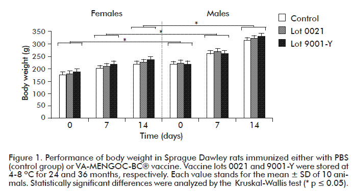
During the study, no clinical symptoms were shown and not any animal died. All the animals gained weight during the 14 days post-inoculation (Figure 1), with no significant differences detected between treatments as compared to the control group (p ≥ 0.05). Male rats gained weight faster than females (p ≤ 0.05), and weight gain curves were in agreement with those previously observed in other experiments, run in our facilities or even reported in the scientific literature [9, 12-16].
Water and food consumption
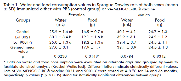
Water and food consumption were similar among evaluated groups (p ≥ 0.05), either receiving treatments or placebo. Males consumed more water and food than females (24.5 g of food and 38.1 mL of water for males vs. 17.9 g and 27 mL females) (Table 1). Mean values were as reported in the literature and consistent with historical records for SD rats in our facilities [7-9, 18, 19]. Nevertheless, significant differences (p ≤ 0.05) were observed in female rats for water consumption between groups immunized with VA-MENGOC-BC® lots 0021 and 9001-Y (stored at 4 to 8 °C for either 24 or 36 months, respectively), and for food consumption between the group receiving lot 0021 and the placebo group. In both cases, consumption was higher in females from the group receiving the vaccine lot 0021. However, these differences were irrelevant from the physiological and toxicological points of view, due to the similarity of weight gain among all the groups and the absence of clinical symptoms. At the same time, the differences seen in female animals immunized with vaccine lot 0021 could be related with a mild fever detected on them at 8 h post-inoculation.
Thermometry and muscular diameter
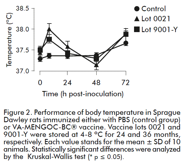
The physiological range of body temperature reported for the rat species is 37.5 ± 0.5 ºC [20]. As shown in figure 2, during the first 8 h there was a slight increase in the mean body temperature in female rats vaccinated with either vaccine lot. Despite average ranges were below 38 ºC, significant differences were found in female rats between for the group receiving the vaccine lot 0021 (37.97 ºC) as compared to placebo (37.43 ºC). In that vaccine group, four out of ten animals had a mild fever (38.1-38.5 ºC), at 8 h post inoculation, which was transient and subsequently disappeared. This could be related to the increased water and food consumption seen in these animals from 8 to 24 h post-inoculation, to recover from the lower consumption rate experimented during the first 8 h when mild fever occurred. Moreover, these parameters were measured every two days. In fact, such effects are well documented in the literature as related to febrile states [21]. Female rats receiving vaccine lot 9001-Y (36 months) showed no significant differences in respect to females of the other two groups. Temperature values at 24, 48 and 72 h post-inoculation were similar (p ≥ 0.05) within physiological ranges.
During the experiment, all the experimental temperature evaluations made on male animals showed mean temperature values similar among groups (p ≥ 0.05).
Importantly, body temperature is determinant for reactogenicity evaluations of vaccine products during clinical trials. In those studies, expected and unexpected adverse events are closely monitored, including: fever, headache, pain at the vaccine administration site, redness, abdominal pain, vomiting and diarrhea, among others [22, 23]. In this sense, thermometry assessment in preclinical toxicological studies can contribute to predict the potential reactogenicity in humans of a given vaccine. Considering this, the results obtained were regarded as satisfactory, with no fever detected in vaccine animals after 8 h post-inoculation, and in agreement with all the other evaluations as evidencing the low reactogenicity of the vaccine tested. They also extended the safety profile of the VA-MENGOC-BC® vaccine.
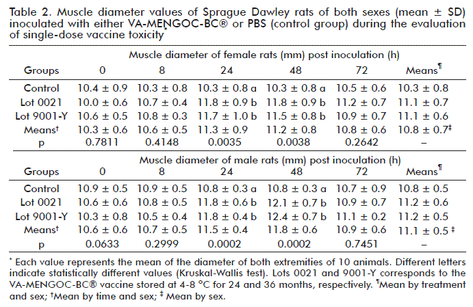
Additionally, the assessment of muscular diameter helps to determine the occurrence of any inflammatory process at the vaccine administration site [24]. This parameter showed significant differences (p ≤ 0.05) in vaccinated animals as compared to the control group at 24 and 48 h, with no significant differences detected between vaccine groups (Table 2). Such an effect was as expected in vaccinated animals due to the aluminum hydroxide present in the formulation as vaccine adjuvant, together with the effect of the immune response against vaccine antigens [13, 25]. Furthermore, muscle diameter peaked at 24 h post-inoculation, with a progression to normal values from 48 h onwards. These was indicative and complemented the low reactogenicity profile of the vaccine, together with the lack of lameness, pain or any other gait dysfunction.
Anatomopathological studies
During necropsies, macroscopic observations were detected at the inoculation site in vaccinated animals. They were consistent with granulomatous processes in both vaccinated groups (Table 3; Figure 3). This type of processes has been consistently reported in animals inoculated with vaccine products containing aluminum phosphate or hydroxide as adjuvant [7, 9, 25]. Moreover, adenitis was present in the popliteal and deep inguinal lymph nodes, possibly related to the immune response activated by the immunogens through the efferent lymph circulation to those ganglia [26]. These effects have been previously reported [7, 9, 25]. No other alterations or macroscopic lesions were found in other organs or organs systems in the animals.
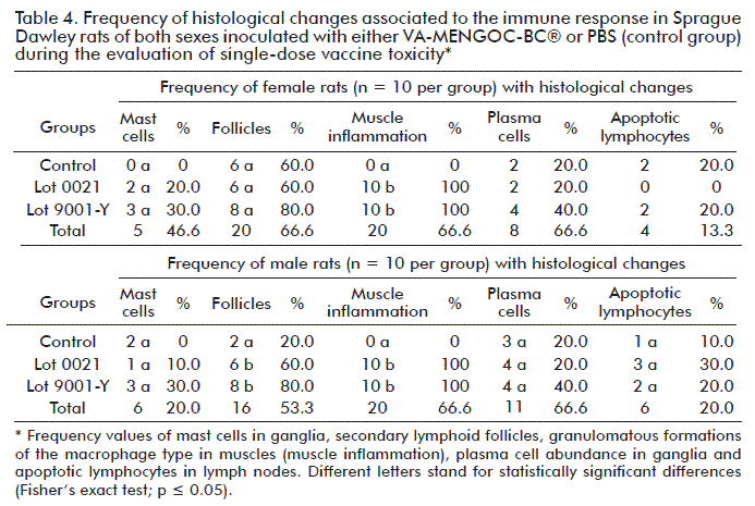
The diagnosed microscopic lesions identified as possible granulomatous processes at the inoculation site were further verified microscopically. Significant differences were found as compared to the inoculation site in the control group (p ≤ 0.05; Table 4). These findings were consistent with observations in similar previous studies carried out in this species and when administering aluminum hydroxide-containing vaccines as VA-MENGOC-BC® [9, 25]. Moreover, the findings in the efferent regional lymph nodes corresponding to the vaccine inoculation site have been regarded as unspecific by other research groups [9, 26]. Despite such considerations, and attending to the significant differences (p ≤ 0.05) observed in vaccinated male rats as compared to the control group in the number of secondary lymphoid follicles in ganglia, it was considered that this effect was related to immunological response against the vaccine.
CONCLUSIONS
In summary, the VA-MENGOC-BC® vaccine stored at 4-8 °C for 36 months showed a similar safety profile in SD rats as that of the vaccine stored for 24 months under the same conditions. Neither local nor systemic adverse toxic effects were detected when a single dose of the vaccine was i.m. administered. This indicates that the vaccine stored for 36 months can be considered as potentially non-toxic and supports the possible extension of VA-MENGOC-BC® vaccine shelf life up to 36 months under these storage conditions.
REFERENCES
1. Batista RS, Gomes AP, Dutra Gazineo JL, Balbino Miguel PS, Santana LA, Oliveira L, et al. Meningococcal disease, a clinical and epidemiological review. Asian Pac J Trop Med. 2017;10(11):1019-29.
2. World Health Organization. Epidemic meningitis control in countries of the African meningitis belt, 2017. Weekly Epidemiological Record. 2018;93(14):173-84.
3. World Health organization. WHO Meeting Report. Developing a new generation RDTs for Meningitis Geneva, 9 March 2018. Geneva: WHO; 2018.
4. WHO. Situation update on meningitis C epidemic risk. Geneva: WHO; 2018.
5. Ochoa RF, Menéndez J. Prevención de la enfermedad meningocócica. La Habana: Ediciones Finlay; 2010.
6. WHO. Guidelines on the nonclinical evaluation of vaccine adjuvants and adjuvanted vaccines Annex 2. WHO Technical Report Series. 2013;(987):1-56.
7. Núñez JF, Herrera L, Infante JF, González P, Pérez V, Argamasilla M, et al. Estudio de toxicidad por dosis única y tolerancia local de una vacuna antimeningocócica tipo B en ratas Sprague Dawley. Vaccimonitor. 2006;15(2):9-14.
8. Fariñas M, Arencibia DF, Días D, Infante JF, Valdés Y, Hernández T, et al. Estudio de toxicidad por dosis única de la vacuna antimeningocóccica ACW135 en ratas Sprague Dawley. Retel. 2011;(35):23-42.
9. Infante Bourzac JF. Estudio de inocuidad e inmunogenicidad protectogénica de la vacuna antimeningocócica VA-MENGOC-BC en modelos murinos. [Tesis Doctoral]. La Habana: Instituto Finlay, Universidad Agraria de La Habana; 2000.
10. Verdier F. Non-clinical vaccine safety assessment. Toxicology. 2002;174(1):37-43.
11. WHO. Guidelines on the nonclinical evaluation of vaccine adjuvants and adjuvanted vaccines. Annex 1. WHO Technical Report Series. 2013;(927):1-36.
12. Lawson T. LATG training manual: laboratory animal technologist. Memphis: America Association for Laboratory Animal Science; 2000.
13. Committee for the Update of the Guide for the Care and Use of Laboratory Animals. Guide for the care and use of laboratory animals. Washington: The National Academies Press; 2011.
14. American Veterinary Medical Association. AVMA Guides for euthanasia of animals. Schaumburg: American Veterinary Medical Association; 2013.
15. Morton DB. Humane endpoints in animal experimentation for biomedical research: Ethical, legal and practical aspects. In: Hendriksen CFM, Morton DB, eds. Humane Endpoints in Animal Experimentation for Biomedical Research. London: Royal Society of Medicine Press; 1999. p. 5-12.
16. Fariñas M, Oliva R, Infante JF, Valdez Y, Nuñez D, Valmaceda T, et al. Ensayo piloto de inmunogenicidad y toxicidad preclínica de la vacuna Salmonella typhi conjugada en ratas Sprague Dawley. Retel. 2014;(44):17-34.
17. Lopez Y, Pastor M, Infante JF, Diaz D, Oliva R, Fernandez S, et al. Repeated dose toxicity study of Vibrio cholerae-loaded gastro-resistant microparticles. J Microencapsul. 2014;31(1):86-92.
18. Charles River Laboratories Inc. SAS Sprague Dawley Rat Details; 2018 [cited 2018 March 27]. Available from: https://www.criver.com/products-services/find-model/sas-sprague-dawley-rat?region=3616
19. Taconic Biosciences. Sprague Dawley® Rat Model. 2018 [cited 2018 March 27]. Available from: https://www.taconic.com/pdfs/sprague-dawley-rat.pdf.
20. Olfert ED, Cross BM, McWilliam AA, editors. Guide to the care and use of experimental animals. Canadian Council on Animal Care. 2nd ed. Ottawa: Bradda Printing Services; 1993.
21. Escobar AL. Fever in children: a critical view of caring practices. Av Enferm. 2017;35(3):333-44.
22. Ochoa RF, Baró IM, Menéndez J, Triana T, Mirabal M, Armesto M, et al. Reactogenicidad e inmunogenicidad de una nueva vacuna de toxoide tetánico y diftérico con concentración reducida en adolescentes cubanos. VacciMonitor. 2006;15(2):13-7.
23. García HM, Thompson R, Valera R, Fando R, Fumane J, Mirabal M, et al. A single dose of live-attenuated 638 Vibrio cholerae oral vaccine is safe and immunogenic in adult volunteers in Mozambique. VacciMonitor. 2011;20(3):1-8.
24. Fariñas M, Oliva R, Hernández T, Hernández M, Nuñez D, Quintero A. Study of reactogenicity of pertussis component adjuved to aluminum hydroxide and aluminum phosphate in Sprague Dawley rats. Avances en Biotecnología Moderna. 2017;S7-P11.
25. Bacardi D, Cosme K, Aldana L, Merino N, Suárez J, Mosqueda O, et al. Preclinical safety testing of the Quimi-Hib® vaccine adjuvanted with aluminum phosphate during product development. Biotecnol Apl. 2013;30(2):118-24.
26. Ahrendt M, Hammerschmidt SI, Pabst O, Pabst R, Bode U. Stromal cells confer lymph node-specific properties by shaping a unique microenvironment influencing local immune responses. J Immunol. 2008;181(3):1898-907.
Received in May, 2018.
Accepted in August, 2018.
Reynaldo Oliva-Hernández. Departamento de Modelos Animales y Toxicología Experimental, Dirección de Investigaciones, Instituto Finlay de Vacunas. Avenida 21, No. 19810 e/ 198 y 200, Reparto Atabey, Playa, La Habana, Cuba. E-mail: roh@finlay.edu.cu; reyolivacuba@gmail.com.

