


My SciELO
 Custom services
Services on Demand
Article
 English (pdf)
 Article in xml format
 Article references
 Send this article by e-mail
Indicators
-
 Cited by SciELO
Related links
-
 Similars in
SciELO
Similars in
SciELO
Share

 Permalink
Cuban Journal of Agricultural Science
On-line version ISSN 2079-3480
Cuban J. Agric. Sci. vol.49 no.3 Mayabeque July.-Sept. 2015
ORIGINAL ARTICLE
Enzymatic hydrolyzed preparation of Saccharomyces cerevisiae: an additive with antibacterial potential for animal feeding
Hidrolizado enzimático de Saccharomyces cerevisiae: un aditivo con potencial antibacteriano para la alimentación animal
Marlen Rodríguez,I Grethel Milián,I Ana J. Rondón,I R. Bocourt,II Marta Laurencio,I Yadileiny Portilla,I A. Beruvides,I
ICentro de Estudios Biotecnológicos, Universidad de Matanzas, Autopista a Varadero, km 3 ½, Matanzas, Cuba.
IIInstituto de Ciencia Animal, Apartado Postal 24, San José de las Lajas. Mayabeque, Cuba.
ABSTRACT
In order to evaluate the antibacterial potential of an enzymatic hydrolyzed preparation of Saccharomyces cerevisiae, three in vitro experiments were performed. The confrontation of the bio-preparation with the pathogenic microorganisms was performed through the methods of substance diffusion in agar, the co-culture establishment and the co-aggregation technique. Bacterial isolations were taken from the liver of sick chicken, and were isolated and identified at the Laboratorio de Investigación y Diagnóstico Aviar (LIDA) in Matanzas. It was demonstrated that the hydrolyzed preparation contains antibacterial substances, mainly bacteriocins and/or antibiotics, which inhibit the growth of Klebsiella spp., Streptococcus spp., Staphylococcus spp., Salmonella spp. and E. coli spp. The antibacterial effect of this additive was demonstrated after decreasing the population of pathogenic bacteria, when they are cultivated in the same medium. The strains of Salmonella spp. and E. coli spp. induce the ability of co-aggregation (32.7 and 22.3 %, respectively) to the components of the cell wall of yeasts within the hydrolyzed preparation. Results indicate the limited growth of bacterial strains in the presence of the enzymatic hydrolyzed preparation of yeast. This activity is multifactorial and unleashes different processes that are interrelated and, as a consequence, favor the improvements of physiology, yield and health of animals. Therefore, it can be used as an additive for animal feeding.
Key words: enzymatic hydrolyzed preparation, antibacterial activity, co-aggregation, pathogenic bacteria.
RESUMEN
Para evaluar el potencial antibacteriano de un hidrolizado enzimático de Saccharomyces cerevisiae, se realizaron tres experimentos in vitro. El enfrentamiento del biopreparado a los microorganismos patógenos se realizó mediante los métodos de difusión de sustancias en agar, el establecimiento de cocultivos y la técnica de la coagregación. Los aislados bacterianos procedían del hígado de pollos enfermos y se aislaron e identificaron en el Laboratorio de Investigación y Diagnóstico Aviar (LIDA) de Matanzas. Se demostró que el hidrolizado contiene sustancias antibacterianas, fundamentalmente bacteriocinas y/o antibióticos que inhiben el crecimiento de Klebsiella spp., Streptococcus spp., Staphylococcus spp., Salmonella spp. y E. coli spp. Se comprobó el efecto antibacteriano de este aditivo, al disminuir la población de bacterias patógenas, cuando se cultivan en el mismo medio. Se observó que las cepas de Salmonella spp. y E. coli spp. inducen habilidad de coagregación (32.7 y 22.3 %, respectivamente) a los componentes de la pared celular de las levaduras presentes en el hidrolizado. Los resultados indican el limitado crecimiento de las cepas bacterianas en presencia del hidrolizado enzimático de levadura. Esta actividad es multifactorial y desencadena diferentes procesos que se interrelacionan y como consecuencia, provocan mejoras en la fisiología, el rendimiento y la salud de los animales. Por ello, se puede inferir su utilización como aditivo en la alimentación animal.
Palabras clave: hidrolizado enzimático, actividad antibacteriana, coagregación, bacterias patógenas.
INTRODUCTION
The biotechnological treatment and use of yeast residues allows the use of food additives for animal production because these are low cost products with positive effects on health and on the zootechnical performance of animals. On the other hand, their utilization contributes to the decrease the risks of environmental contamination (Fleet 2007, Jacques and Casaregola 2008 and Pérez et al. 2012).
Among the substances with prebiotic activity, there are some of the structural components of the cell walls of yeasts, and the strains from Saccharomyces cerevisiae are among the most used. Polysaccharides and other products of their hydrolysis produce prebiotic effects, mainly because they stimulate the immunological response and prevent infectious diseases in animals and humans (Lourenco et al. 2011and Van-Staden and Dicks 2012).
Because of its nutritional, pharmacological and dynamic characteristics, the strains of Saccharomyces cerevisiae (Sc) have been used in animal feeding for several decades, because they provide proteins, minerals, and B complex vitamins. In addition, their cell wall is formed, partly, by mannanes and beta-glucanes that, after being released, may favor the intestinal exclusion of pathogenic bacteria (Moslehi-Jenabian et al. 2010 and Carro et al. 2014).
Pérez et al. (2006) described the methodology for obtaining the enzymatic hydrolyzed preparation of Saccharomyces cerevisiae. This product contains viable cells of Bacillus and its endospores, which potentiate the favorable effects of this bio-preparation on the prevention of infectious diseases in animals of zootechnical importance (Pérez et al. 2011 and Milián et al. 2014). However, it is not known which specific antimicrobial action develops this bio-preparation in front of harmful bacteria from the gastrointestinal ecosystem. Therefore, the objective of this study was to evaluate the antibacterial potential of an enzymatic hydrolyzed preparation of Saccharomyces cerevisiae in front of potentially pathogenic strains, using three in vitro experiments.
MATERIALS AND METHODS
Production of an Enzymatic Hydrolyzed Preparation of Yeast. The cream of Saccharomyces cerevisiae was obtained from an alcohol distillery plant belonging to the Complejo Agroindustrial “Jesús Rabí” in Calimete, Matanzas province. For preparing the enzymatic raw and producing the hydrolyzed of S. cerevisiae yeast, the methodology described and registered by Pérez et al. (2006) was used.
Treatment of the indicator strains. Wild strains of Klebsiella spp., Streptococcus spp., Staphylococcus spp., Salmonella spp. and E. coli were used, which were isolated from the liver of sick fowls and identified at the Laboratorio de Investigación y Diagnóstico Aviar (LIDA) from Matanzas. All the indicator strains were inoculated in a nutrient-enriched medium and incubated into a shaker (MAXQ-600), under anaerobic conditions for 24 hours at 37 ºC.
Determination of the antibacterial effect of the hydrolyzed preparation in co-cultures with potentially pathogenic bacteria. The antagonistic activity of an enzymatic hydrolyzed preparation of yeast was studied through the associated cultures or culture mixtures. The technique described by Orlowski and Bielecka (2006), and modified by Rodríguez (2010), was used. The development of a co-culture took place in a shiny green bile medium (BVB, Biocen), which was added in flasks of 100 mL, at a rate of 50 mL of effective volume. An amount of 3.6 mL of the enzymatic hydrolyzed preparation and 5 mL of the culture with the potentially pathogenic or indicator microorganism (with a population of 1x10⁹ CFU.mL-1) were added to each flask. Once they are mixed, the co-cultures were incubated under static conditions for 12 h at 37 ºC, and samples were taken at 0, 4, 8, 12, 16, 20 and 24 h in order to perform serial dilutions in a peptone medium (1 %). The culture took place in plaques with Agar MacConkey (Biocen) for counting viables. As indicator strains, the pure strains of Klebsiella spp., Streptococcus spp., Staphylococcus spp., Salmonella spp. and E. coli were used, which were inoculated in the same medium (BVB).
Determination of antibacterial substances. The method of diffusion of substances in agar was applied, which was proposed by Schillinger and Lucke (1989).
Determination of antibacterial substances of the hydrolyzed preparation in front of positive and negative Gram bacteria. A total of 10 mL of the hydrolyzed preparation were taken and they were centrifuged at 15,000 rpm at 5 ºC for 10 min. in a refrigerated centrifuge (P-selecta-Mixtasel). Later, the supernatant was sterilized with cellulose acetate filters, with pores of 0.22 μm (Minisart, satorius 600 kPa max). The supernatant was used in three variants: V1- supernatant without modification, V2 – modified supernatant, with the addition of NaOH 0.1 N (up to pH 7) to remove the action of acids, and V3 – modified supernatant with the addition of the pronase enzyme (1 mg. mL-1) to remove the action of bacteriocins. The potentially pathogenic strains used in the previous experiment were used as indicator strains.
Development of the technique of diffusion in agar. An amount of 200 μL was taken from the cultures of the indicator strains. They were inoculated in tubes with 20 mL of nutrient enriched agar (with 1 % of Ion-Agar, OXOID) at 45 ºC. They were poured in plaques for their solidification. In each plaque that contained indicator strains, small wells of 7 mm of diameter were opened, in which 20 μL of variants 1, 2 and 3 of the supernatant of the hydrolyzed preparation were deposited. The plaques were maintained at 5 ºC for 4 h until achieving the best diffusion of substances in agar. Later, they were incubated for 24 h at 37 ºC until finding the growth and appearance of inhibition halos. The diameter of halos was measured with a ruler. The diameter of the small wells was subtracted from each value.
Determination of the co-aggregation of potentially pathogenic bacteria to the components of the cell Wall of yeasts within the the hydrolyzed preparation. The percent of co-aggregation was determined using the formula of Orłowski and Bielecka (2006).
Percent of co-aggregation={[(AXt +Ayt)/2 – At (x + y) / (Axi + Ayi)/2)]}. 100
Where:
AXt: Absorbency at 5 h of the indicator strain
Ayt: Absorbency at 5 h of the yeast hydrolyzed preparation
At (x + y): Absorbency of the mixture of indicator strain + yeast hydrolyzed preparation
Axi: Absorbency of the indicator strain in the initial time
Ayi: Absorbency of the yeast hydrolyzed preparation in the initial time
Statistical processing of data. Three replications were performed per each experiment and per each simple. The results were analyzed according to a design of simple classification. The system INFOSTAT Version 1 (Balzarini et al. 2001) was used for data processing. The analysis of variance were performed to verify the differences among means, with a level of significance of P<0.05. The test of Duncan (1955) was used to perform multiple comparisons among the means. The counting of viable microorganisms was turned into Log N to guarantee the normality conditions in the growth curve.
RESULTS AND DISCUSSION
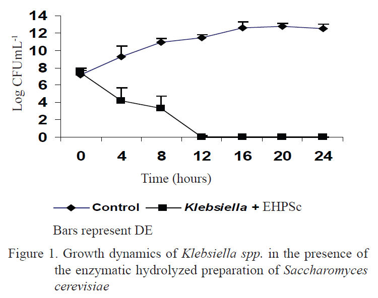
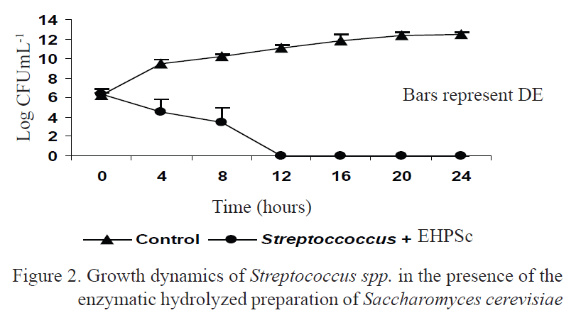
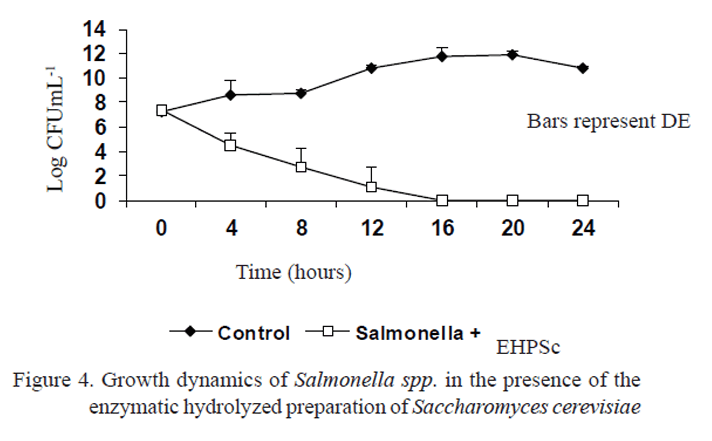
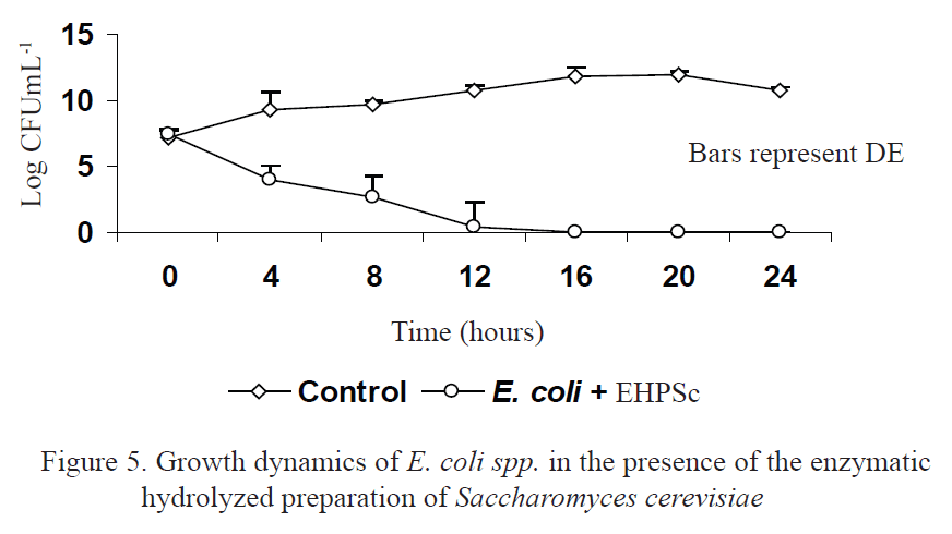
Figures 1, 2, 3, 4 and 5 show the results of the development of co-cultures with the enzymatic hydrolyzed preparation and the indicator strains Klebsiella spp., Streptococcus spp., Staphylococcus spp., Salmonella spp. and E. coli. There was no growth of the indicator strains in the medium with the dose used in this bio-preparation, while they showed an accelerate growth in the control culture.
It was verified that, as the time went by, the cells of indicator strains lost their viability and, after 12 hours, there were no live cells in the co-cultures of Klebsiella spp., Streptococcus spp., Staphylococcus spp. and E. coli spp. The co-culture of Salmonella spp. showed a total reduction after 16 hours. These results indicate that there are antimicrobial substances in this bio-preparation that limit the growth of the inoculated pathogenic strains.
It is known that hydrolysis of the cell wall components of Saccharomyces cerevisiae is achieved using enzymes that produce Bacillus cells. The literature refers to the function of this genus in the production of bactericide and bacteriostatic substances (Milián 2009 and Ayala et al. 2012).
Similar results have been reported by Svetoch et al. (2011), and Bayoda (2013), who demonstrate the antagonistic activity of a hydrolyzed preparation of Saccharomyces cerevisiae and of Bacillus subtilis 21BMC, respectively, in front of different pathogenic microorganism, including the Candida albicans, Proteus, E. coli, Shigella and Salmonella.
According to Cho et al. (2011), the utilization of bacteria from Bacillus genus is a result of their ability to produce endospores. Studies carried out by Leser et al. (2008) state that these strains (spores or vegetative cells) exclude pathogenic microorganisms by competitive adhesion or by synthesis of antimicrobial substances.
The presence of Bacillus and its endospores in the hydrolyzed preparation of yeast could cause the inhibition of the growth of indicator strains in front of this bio-preparation.
Table 1 shows the results of the determination of the antibacterial substances in the enzymatic hydrolyzed preparation in front of indicator strains.
The enzymatic hydrolyzed preparation of yeast caused the formation of inhibition halos in the supernatants of V1 and V2 in front of strains of Klebsiella spp., Streptococcus spp., Staphylococcus spp., Salmonella spp. and E. coli spp. These results indicate the presence of bacteriocins and/or antibiotics in this hydrolyzed preparation, because there was an inhibition of the indicator strains with the supernatant of V3 (acid production).
Similar studies, carried out by Vondruskova et al. (2010) and Ansari et al. (2012), demonstrate that different strains of Bacillus subtilis are able of generating substances like bacitracin, polymixin, difficidin, subtilin and mycobacillin, which are antibiotics that inhibit the growth of pathogenic microorganisms from the intestine of animals.
The inhibiting activity of Bacillus subtilis was also evaluated by mass spectroscopy (Lim-Teo et al. 2005) and it was demonstrated that this bacteria does not have the ability to inhibit Clostridium difficile, Streptococcus pneumoniae, Campylobacter jejuni ATCC-35918, Campylobacter coli ATCC-51798 and Clostridium perfringens ATCC- 13124, due to the production of bacteriocins.
According to these results, the highest inhibiting activity existed in front of the strain of Salmonella (P<0.05), followed by E. coli. In young animals, 70 % of the infections due to enterobacteria show the presence of these agents as the most commonly isolated microorganisms (Pérez et al. 2011 and Tellez et al. 2012).
According to the literature (Gusils et al. 2008 and Fernando 2008), Gram-negative bacteria, like E. coli and Salmonella, use fimbriae to adhere to the target cells of the intestine of animals. These proteins (adhesins), within the fimbriae of pathogenic microorganisms, fix themselves to the receptors of cell walls of yeasts, and are capable of dragging pathogenic bacteria, due to their ability of bonding these microbes to their cell walls or joining by the receptive areas of the pathogens, which are connected to the intestinal mucus (Pérez-Sotelo et al. 2005 and Moslehi-Jenabian et al. 2010).
During the last years, the use of probiotics in prophylaxis and therapy of gastrointestinal diseases has been a subject of great interest and scientific discussion. Nowadays, the importance and possible efficiency of biotic therapy (probiotics and prebiotics) has been recognized as a medical tool for treating digestive diseases (Carro et al. 2014).
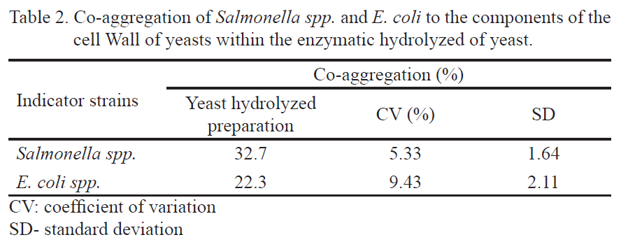
Table 2 shows the percent of co-aggregation reached by the cells of Salmonella spp. and E. coli spp. to the components of the cell wall of yeasts within the enzymatic hydrolyzed preparation of S. cerevisiae.
According to Suman Saran et al. (2012), the self-aggregation in bacteria can be defined as the phenomenon of aggregation among cells of the same strain, while the co-aggregation means the aggregation occurring between different species. The capacity of aggregation is related to the adhesion, which is a characteristic of E. coli and Salmonella spp.
As table 2 shows, the percentages of co-aggregation reported in this study could be related to the significant presence of cell wall fractions of yeasts within the enzymatic hydrolyzed preparation. Regarding this fact, Sánchez-Hernández et al. (2012) stated that when pathogens add to the cell wall of yeasts, a protective effect is induced, and this S. cerevisiae-pathogen complex is rapidly removed from the digestive tract.
Scientific studies demonstrate that mannane oligosaccharides (MOS) are efficient for the agglutination of pathogens like Salmonella and E. coli, regulate the colonization of pathogens and generate the reestablishment of the beneficial flora, improving the intestinal health of animals (Celyk et al. 2003 and Khati et al. 2007). The adhesion property of MOS is demonstrated in this experiment and reveals the probiotic potential of this additive in antimicrobial activity.
For several years, multidisciplinary groups have worked on the introduction of these bio-preparations in animal production, due to their effect on yield and health of animals, and to the use of an economically viable technology for Cuban conditions (Pérez et al. 2006).
The results achieved in this study demonstrate the antibacterial potential of the enzymatic hydrolyzed preparation of yeast in front of the evaluated pathogenic microorganisms. The inhibiting activity, due to the production of bacteriocins and/or antibiotics and to the co-aggregation with pathogens, indicates the limited growth of bacterial strains in the presence of this bio-preparation. This activity is multifactorial and unleashes different processes that are interrelated and provoke, as a consequence, the improvements of physiology, yield and health of animals. Therefore, it can be used as an additive for animal feeding.
REFERENCES
Ansari, A., Aman, A., Siddiqui, N., Iqbal, S., Ali, U. & Qader, S. 2012. ‘‘Bacteriocin (BACIB17): screening, isolation and production from Bacillus subtilis KIBGE IB-17’’. The Karachi Institute of Biotechnology & Genetic, 25: 195.
Ayala, L., Bocourt, R., Milián, G., Castro, M., Herrera, M. & Guzmán, J. 2012. ‘‘Assessment of a probiotic based on Bacillus subtilis and its endospores in the obtainment of healthy lungs of pigs’’. Cuban J. Agric. Sci., 46: 391.
Bayoda, B. 2013. Evaluación in vitro de la actividad antimicrobiana de una cepa de Bacillus subtilis con potencial probiótico. Engineering Thesis, Universidad de Matanzas, Matanzas, Cuba.
Carro, M. D., Saro, S., Mateos, I., Días, A. & Ranilla, M. J.n.d. Perspectivas y retos de los extractos vegetales como aditivos alimentarios en rumiantes. Grupo Asís Biomedia, S.L., 4-6 p.
Celyk, K., Denly, M. & Savas, T. 2003. ‘‘Reduction of toxic effects of Aflatoxin B1 by using Baker yeast (S. cerevisiae) in growing broiler chicken diets’’. Rev. Bras. Zootec, 32: 615.
Cho, J. H., Zhao, P. Y. & Kim, I. H. 2011. ‘‘Probiotics as a Dietary Additive for Pigs. J. Animal and Veterinary Advances’’. In: vol. 10, p. 2127.
Duncan, B. 1955. ‘‘Multiple ranges and multiple F test’’. Biometrics, 11: 1.
Fernando, N. 2008. Utilización de oligosacáridos mananos como promotor de crecimiento en cría y levante de pollitas de reposición Lohman Brown y su efecto hasta el pico de producción. Engineering Thesis, Escuela Superior Politécnica de Chimborazo, Chimborazo. Ecuador.
Fleet, G. H. 2007. ‘‘Yeasts in foods and beverages: impact on product quality and safety’’. Curr. Opinion Biotechnol, 18: 170.
Gusils, C., Oppezzo, O., Pizarro, R. & Gonzalez, S. 2008. ‘‘Adhesion of probiotic lactobacilli to chick intestinal mucus’’. Can. J. Microbiol, 49: 472.
INFOSTAT 2009. Grupo InfoStat, FCA. version 1, Universidad Nacional de Córdova, Argentina.
Jacques, N. & Casaregola, S. 2008. ‘‘Safety assessment of dairy microorganisms: The hemiascomycetous yeasts’’. Int. J. Food Microbiol, 126: 321.
Khati, B., Kolte, B., Shendare, R., Palve, H., Mandlekar, S. & Shisodiya, J. 2007. ‘‘Effect of low protein level supplemented with or without yeast (Saccharomyces cerevisiae) on haematological and immunological profile of broiler quails’’. Royal Veterinary J. of India, 3: 131.
Leser, T. D., Knarreborg, A. & Worm, J. 2008. ‘‘Germination and outgrowth of Bacillus subtilis and Bacillus licheniformis spores in the gastrointestinal tract of pigs’’. J. Appl. Microbiol, 104: 1025.
Lim, T., Yeow, A. & Tan, M. 2005. ‘‘Inhibition of Clostridium perfringens by a Novel Strain of Bacillus subtilis Isolated from the Gastrointestinal Tracts of Healthy Chickens’’. Appl. Environmental Microbiol, 71: 4185.
Lourenço, M., Hayashi, R. M., Pickler, L., Miglino, L. B., Kuritza, L. & Santin, E. 2011. ‘‘Expresión de células caliciformes y linfócitos t cd3 en la mucosa intestinal de pollos de engorde suplementados con oligosacáridos mananos’’. In: XXII Congreso Latinoamericano de Avicultura, Curitiba, Brasil: Universidad Federal de Paraná.
Milián, G. 2009. Obtención de cultivos de Bacillus spp. y sus endosporas. Evaluación de su actividad probiótica en pollos (Gallus gallus domesticus). PhD Thesis, Instituto de Ciencia Animal, La Habana, Cuba.
Milián, G., Rondón, A. J., Pérez, M., Samaniego, L. M., Riaño, J., Bocourt, R., Ranilla, M. J., Carro, M. D., Rodríguez, M. & Laurencio, M. 2014. ‘‘Isolation and identification of strains of Bacillus spp. in different ecosystems, with probiotic purposes, and their use in animals’’. Cuban J. Agric. Sci, 48: 347.
Moslehi-Jenabian, S., Lindegaard, L. & Jespersen, L. 2010. ‘‘Review: beneficial effects of probiotic and food borne yeasts on human health’’. Nutrients, 2: 449.
Orłowski, A. & Bielecka, M. 2006. ‘‘Preliminary characteristics of Lactobacillus and Bifidobacterium strains as probiotic candidates’’. Pol. J. Food Nutr. Sci., 15: 269.
Pérez, M., Laurencio, M., Rondón, A. J., Milián, G., Arteaga, F., Rodríguez, M. & Borges, Y. 2012. ‘‘Evaluación de una mezcla probiótica en la alimentación de gallinas ponedoras en una unidad de producción comercial’’. Rev. Pastos y Forrajes, 35: 311.
Pérez, M., Laurencio, M., Rondón, A. j, Milián, G., Bocourt, R. & Arteaga, F. 2011. ‘‘Actividad antimicrobiana de una mezcla probiótica de exclusión competitiva y su estabilidad en el tiempo’’. Rev. Salud Anim., 33: 147.
Pérez, M. Q., Milián, G., Piad, R. B., González, R. C., Bocourt, R. S. & Savón, V. 2006. Hidrolizado de fondaje de cubetas de destilerías de alcohol con un crudo enzimático de la cepa de Bacillus licheniformis E-44 y su procedimiento de obtención. no. 23179. (Int.cl.8) A 23 J 1/00, 3/30, C 12N 9/56.
Pérez-Sotelo, L. S., Talavera, R. M., Monroy, H. G., Bernabé, S. L., Cuarón, J. A., Ibargüengoytia, R., Montes de Oca, J. & Vázquez, J. C. 2005. ‘‘In vitro evaluation of the binding capacity of Saccharomyces cerevisiae Sc47 to adhere to the wall of Salmonella spp’’. Revista Latinoamericana de Microbiología, 47: 70.
Rodríguez, M. 2010. Evaluación in vitro de la actividad antimicrobiana del hidrolizado enzimático de levadura Saccharomyces cerevisiae. Master Thesis, Instituto de Ciencia Animal, Cuba.
Sánchez-Hernández, J. A., Mayta-Baldivieso, M. J. & Rivera-Tapia, J. A. 2012. ‘‘Alteraciones del pH vaginal asociado a lactobacilos o bacilo de Doderlein’’. Revista Latinoamericana de Patología Clínica y Medicina, 59: 56.
Schillinger, U. & Lucke, F. K. 1989. ‘‘Antibacterial Activity of Lactobacillus sake Isolated from Meat’’. Appl. Environm. Microbiol., 55: 1901.
Suman Saran, M. S., Bisht, K., Singh, U. V. & Teotia, S. 2012. ‘‘Comparing Adhesion Attributes of two Isolotes of Lactobacillus Acidophilus for Assessment of prebiotics Honey and Inulin’’. International J. Sci. Research Publications.
Svetoch, E. A., Eruslanov, B. V., Levchuk, V. P., Perelygin, V. V., Mitsevich, E. V., Mitsevich, I. P., Stepanshin, J., Dyatlov, I., Seal, B. S. & Stern, N. J. 2011. ‘‘Isolation of Lactobacillus salivarius 1077 (NRRL B-50053) and Characterization of Its Bacteriocin, Including the Antimicrobial Activity Spectrum’’. Appl. Environ Microbiol, 77: 2749.
Tellez, G., Pixley, B. E., Wolfenden, S., Layton, L. & Hargis, B. 2012. ‘‘Probiotics/direct fed microbials for Salmonella control in poultry G Probiotics/direct fed microbials for Salmonella control in poultry’’. Food Research International, 45: 628.
Van Staden, A. D. & Dicks, L. M. 2012. ‘‘Applications, antibiotic release and alternatives to antibiotics’’. J Appl. Biomater Funct Mater, 10: 2.
Vondruskova, H., Slamova, R., Trckova, M., Zraly, Z. & Pavlik, I. 2010. ‘‘Alternatives to antibiotic growth promoters in prevention of diarrhea in weaned piglets: a review’’. Veterinarni Medicina, 55: 199.
Received: February 2, 2015
Accepted: June 22, 2015
Marlen Rodríguez, Centro de Estudios Biotecnológicos, Universidad de Matanzas, Autopista a Varadero, km 3 ½, Matanzas, Cuba. Email: marlen.rodriguez@umcc.cu

