


My SciELO
 Custom services
Services on Demand
Article
 English (pdf)
 Article in xml format
 Article references
 Send this article by e-mail
Indicators
-
 Cited by SciELO
Related links
-
 Similars in
SciELO
Similars in
SciELO
Share

 Permalink
Cuban Journal of Agricultural Science
On-line version ISSN 2079-3480
Cuban J. Agric. Sci. vol.49 no.4 Mayabeque Oct.-Dec. 2015
ORIGINAL ARTICLE
Optimization of an immunoenzymatic (ELISA) assay for detecting ovine antibodies against Haemonchus contortus
Optimización de un ensayo inmunoenzimático (ELISA) para detectar anticuerpos de ovinos contra Haemonchus contortus
A. Díaz,I A. Arenal,I Jessea França,II Alda L. Gomes,III Maria A. Machado,III M. Sossanovicz,II Úrsula Yoshitani,II M.B. Molento,II
ILaboratorio de Bioquímica, Departamento de Morfofisiología, Universidad de Camagüey km 5 ½ 74650, Cuba.
IILaboratorio de Enfermedades Parasitarias, Universidad Federal de Paraná, Curitiba, Brasil, Rua dos Funcionários, 1540. Curitiba, PR, CEP: 80.035-050.
IIIDepartamento de Zootecnia, Universidad Federal de Paraná, Curitiba, Brasil, Rua dos Funcionários, 1540. Curitiba, PR, CEP: 80035-050.
*Estos autores contribuyeron por igual a la realización del trabajo. Email: amilcar.arenal@reduc.edu.cu.
ABSTRACT
Diseases originated by gastrointestinal nematodes are one of the most important causes of economic losses in animals, mainly in small ruminants. The objective of this study was to optimize an ELISA immunoenzymatic assay for Haemonchus contortus. For that, an extract of total proteins was extracted from a macerate of the adults of this nematode. Blood samples were taken of 48 sheep for obtaining the serum. The parameters of an immunoenzimatic (ELISA) assay were optimized. The best concentration for covering the antigen was 5 µg/mL. The best relationships between the signals of the positive and negative serums for serum dilution, the Anti-IgG and the bovine fetal serum were obtained in 1:300, 1:7000 and 0.25 %, respectively. The optimization of the parameters of this ELISA immunoenzymatic assay allowed the detection of IgG that recognize Haemonchus contortus with low unspecific interactions. This is the first study available on the optimization of an ELISA assay for Haemonchus contortus.
Key words: optical density, IgG, Haemonchus contortus.
RESUMEN
Las enfermedades ocasionadas por nemátodos gastrointestinales constituyen una de las más importantes causas de pérdidas económica en los animales, fundamentalmente en los pequeños rumiantes. El objetivo de este estudio fue optimizar un ensayo inmunoenzimático de ELISA para Haemonchus contortus. Para ello, se obtuvo un extracto de proteínas totales a partir de un macerado de los adultos de este nemátodo. Se tomaron muestras de sangre de 48 ovinos para obtener el suero. Se optimizaron los indicadores de un ensayo inmunoenzimático (ELISA). La mejor concentración para el recubrimiento del antígeno fue de 5 µg/mL. Las mejores relaciones entre las señales de los sueros positivos y negativos para la dilución de los sueros, el Anti-IgG y el suero fetal bovino se obtuvieron en 1:300. 1:7000 y 0.25 %, respectivamente. La optimización de los parámetros de este ensayo inmunoenzimático de ELISA permitió la detección de IgG que reconocen Haemonchus contortus, con bajas interacciones inespecíficas. Este es el primer trabajo que se tiene reporte sobre la optimización de un ensayo de ELISA para Haemonchus contortus.
Palabras clave: densidad óptica, IgG, Haemonchus contortus.
INTRODUCTION
Gastrointestinal nematodes (GIN) are one of the most important causes of economic losses for ruminant exploitations. They generate weight gain reduction, growth delay, decrease of feed intake, reduction of milk yield, wool and low fertility and, in cases of mass infections, animal death (Kui-zheng et al. 2007, Liu et al. 2009 and Oliveira et al. 2012). From the GIN, the most important for small ruminants is Haemonchus contortus, which is present in warm climates (Giudici et al. 1999, Amarante 2014 and Wilmsen et al. 2014) and in temperate regions (Waller et al. 2004, Mederos et al. 2010 and Felippelli et al. 2014).
For Haemonchus contortus diagnosis, the most utilized method consists of techniques for determining the presence and/or number of parasite eggs (Ward et al. 1997). Within these techniques, McMaster is the most worldwide popular (Bosco 2014) and more recently the Mini-FLOTAC (Rinaldi 2014). Post-mortem examination (necropsy) in slaughter houses allows the confirmation of the diagnosis through the finding of the adult parasites (Sissay et al. 2007, Qamar et al. 2009 and Khalafalla et al. 2011).
The ELISA immunoenzymatic assay is one of the most used tools for establishing diagnostic methodologies for the most dissimilar parasites both in humans as in animals: Taenia solium (Deplazes et al. 1991), Fasciola hepatica (Howell et al. 2015), Crypto sporidium (Elgun and Koltas 2011), Schistosoma mansoni (El-Badry 2009), Trichinella spiralis (Johnson et al. 1996) and Taenia saginata (Wandra et al. 2006). However, for Haemonchus contortus there is still no immunoenzymatic method allowing animal diagnosis.
The objective of this study was to optimize the ELISA immunoenzymatic assay for Haemonchus contortus.
MATERIALS AND METHODS
Haemonchus contortus proteins extract. Adult H. contortus, collected from the abomasums of slaughtered animals were macerated in 10 g liquid nitrogen and 10 mL of saline solution buffered with phosphates (SSTP) 1 X (KH2PO4 1.8 mmol/L; Na2HPO4 8.4 mmol/L; KCl 12.6 mmol/L and NaCl 136.8 mmol/L, pH 7.2). Later all this macerated material was deposited in a 50 mL centrifuge test tube and it was adjusted until 20 mL with SSTP IX. The tube was agitated for 30 min at 4 ºC. Afterwards it was centrifuged at 10000 rpm for 20 min at
4 ºC. The supernatant was collected and filtered. Protein determination was carried out through the QuantiProTM High Sensitivity Protein Assay Kit.
Sampling. For establishing ELISA, samples of blood serum were used of 48 female sheep from the Laboratory of Sheep and Goat Research and Production (LAPOC) belonging to the Federal University of Paraná in Brazil. To all animals fecal feces were taken for realizing the modified method of McMaster and coproculture. In this way the infestation or not by the nematode was determined. Blood obtained was centrifuged at 3000 rpm for 10 min. A mixture with similar volumes of all serums was conformed and stored at -20º C.
Immunoenzymatic (ELISA) assay. Microtitration plates of 96 dishes (PolySorp, Nunc) were recovering the concentration of 5 µg/mL (100 µL/dish) with the Haemonchus contortus proteins extract diluted in recovering buffer (carbonate-sodium bicarbonate 0.1 mM, pH 9.6) and incubated at 4º C throughout the night. Dishes were washed three times with SSTP IX solution with Tween 20 at 0.2 % ([vol/vol]). The blocking was made with Bovine Fetal Serum (BFS) at 3 % ([vol/vol]) diluted in SSTP 1X with Tween 20 at 0.2 % for one hour. Dishes were washed three times with SSTP 1X solution with Tween 20 at 0.2 % [vol/vol]. Serum samples of vaccinated animals with the parasite extract and the controls were applied (100 µL/dish) diluted in SSTP IX with Tween 20 at 0.2 % ([vol/vol]) and BFS at 25 % ([vol/vol]). After plate incubation for one hour at environmental temperature, dishes were washed three times with SSTP IX solution with Tween 20 at 0.2 % [vol/vol] and the Anti IgG of sheep was added diluted 1:7000 in SSTP IX with Tween 20 at 0.2 % ([vol/vol]) and BFS at 0.25 % ([vol/vol]) was incubated for one hour. The dishes were washed three times with SSTP IX solution with Tween 20 at 0.2 % ([vol/vol]) and the buffer ABTS (revealing) was added, it was incubated in the darkness for 30 min. The optical density was determined in the Biotek reader EL x 800 at 405 nm.
Statistical analysis. For data processing the statistical package Graph Pad Prism 5 was used. An analysis of variance (ANOVA) was carried out and for mean comparison the Post hoc test of Student Newman-Keuls was used.
RESULTS AND DISCUSSION
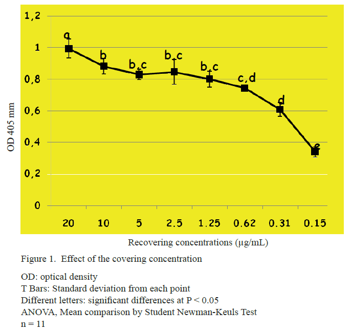
In figure 1 is shown the effect of the recovering at different antigen concentrations. A better performance between 20 and 5 µg/mL is observed with values of optical density of one for the first and 0.82 for the second. For the rest, the signal decreases until attaining the minimum concentration assessed (0.15 µg/mL) with optical density value close to 0.3.
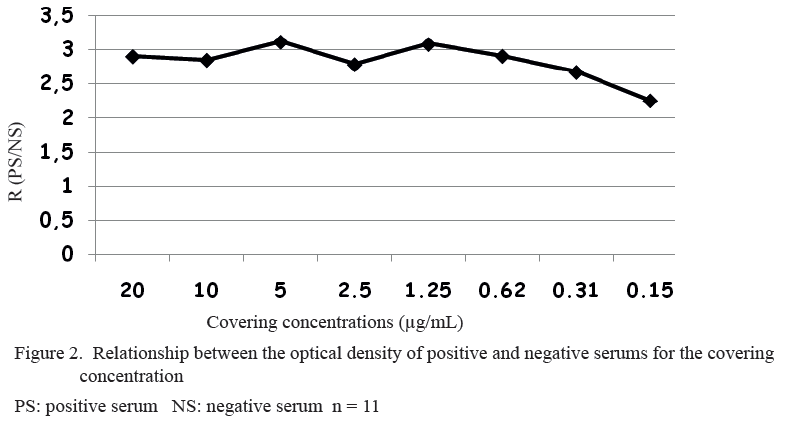
The best relationship between the optical density of the positive and the negative serums (figure 2) were found in 5 µg/mL of the covering concentration. Other reports used the same covering concentration for the detection of titers against Haemonchus contortus (Domínguez- Toraño et al. 2003, Shakya et al. 2011 and Fawzi et al. 2014). Nonetheless, coverings of 1 and 2 µg/mL were employed for the determination of antibodies induced by vaccination with this nematode (Bakker 2004, De Vries et al. 2009, Bassetto et al. 2011 and Molina et al. 2012). Meanwhile the covering with 10 µg/mL was also used (van Stijn et al. 2010).
Figure 3 shows the effect of the dilution of the animal serum. The 1:200 dilution was of the highest optical density (1.1). The remaining dilutions behaved very similar but with a tendency to the decrease of the signal.
The relationship between the optical density of the positive and negative serums is shown in figure 4. In the 1:300 dilution 2.7 relationship was obtained which is much higher to that of 1:200 that was of 2.2. Reports indicate the utilization of serums for detecting antibodies against Haemonchus contortus at the most different concentrations, 1:100 (De Vries et al. 2009); 1:200 (Ruiz et al. 2004, Bassetto et al. 2011 and Molina et al. 2012); 1:1000 (Sun et al. 2011, Han et al. 2012 and Yan et al. 2013) and 1:4000 (Smith et al. 2003).
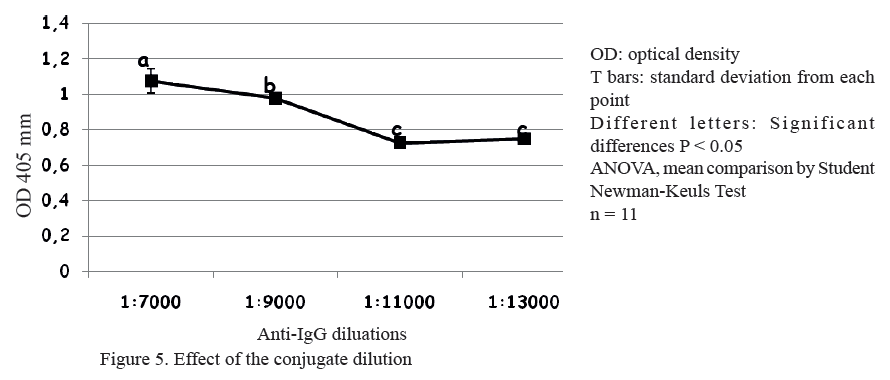
The effect of the conjugate (Anti-IgG of sheep) dilution is shown in figure 5. The conjugate dilution recommended by the producer (Invitrogen®, USA) was of 1:7000. Nonetheless, conjugate dilutions from 1:7000 to 1:13000 were evaluated. The dilutions 1:7000 and 1:9000 exhibited values of optical densities higher to one. On diluting more the conjugate the tendency was to decrease the signal with minimum in 1:11000 and 1:13000.
Figure 6 presents the relationship between the optical density of the positive and negative serums. The conjugate dilution 1:7000 was that of highest relationship. Some authors report the utilization of the conjugate at a dilution of 1:8000 (Fawzi et al. 2014) which is similar to ours. On the contrary, in other reports conjugate dilutions ranging from 1:3000 (Bakker 2004), 1:4000 (Ruiz et al. 2004 and Molina et al. 2012), 1:5000 (Santos et al. 2014), 1:10000 (Bassetto et al. 2011), 1:20000 (Muleke et al. 2007) up to 1:25000 (Shakya et al. 2011).
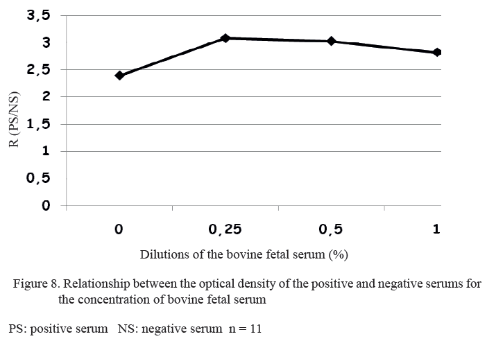
For serum incubation and the conjugate (Anti-IgG), BFS was used for decreasing the unspecific interactions of the assay. For this, dilutions were assessed from 0 until 1 % (figure 7). In the dilutions where no BFA was utilized and in that of 0.25 %, the values of optical density were approximately one. As BFS concentration increased, the tendency was to decrease the values of optical density.
Figure 8 shows the relationship between the positive and negative serums for determining BFS concentration. The highest relationship was attained with the dilution at 0.25 %. When BFS concentration was increased, the signal decreased. BFS dilutions at 1 % (Rodríguez Sánchez et al. 2005) and at 2 % (Pupo-Antúnez et al. 2011) were employed with the purpose of eliminating the unspecific interactions in ELISA assays. Bashir et al. (2011) and Wu et al. (2015) reported the use of albumin from bovine serum for this objective. The first do not declare the concentration utilized while the second made it at 1 %.
The optimization of the parameters of this ELISA immunoenzymatic assay allowed achieving high optical density values with low unspecific interactions. With this procedure the effect of Haemonchus contortus can be assessed on meat and wool production without the need of animal slaughtering. For this, it is necessary the identification of the gray zone of this assay.
ACKNOWLEDGEMENTS
The authors are indebted to CAPES and BIOLAC for financing pre-doctoral scholarships that allowed the development of this research. To the Federal University of Paraná (UFPR), to all its technicians, staff members, students and professors for their inestimable help.
REFERENCES
Amarante, A. F. T. 2014. “Sustainable worm control practices in South America”. Small Ruminant Research, 118 (1): 56–62.
Bakker, N., Vervelde, L., Kanobana, K., Knox, D. P., Cornelissen, A., De Vries, E. & Yatsuda, A. P. 2004. “Vaccination against the nematode Haemonchus contortus with a thiol-binding fraction from the excretory/secretory products (ES)”. Vaccine, 22 (5): 618–628.
Bashir, S., Singh, R., Sharma, B. & Yadav, S. K. 2011. “Development of a sandwich ELISA for the detection of bovine herpesvirus type 1”. Asian Pacific J. Tropical Medicine, 4: 363.
Bassetto, C. C., Silva, B. F., Newlands, G. F. J., Smith, W. D. & Amarante, A. F. T. do 2011. “Protection of calves against Haemonchus placei and Haemonchus contortus after immunization with gut membrane proteins from H. contortus”. Parasite immunology, 33 (7): 377–381.
Bosco, A. 2014. The coprological diagnosis of gastrointestinal nematode infections in small ruminants. Ph.D. Thesis, Università degli Studi di Napoli Federico II, Napoli, Italia.
Deplazes, P., Eckert, J., Pawlowski, Z. S., Machowska, L. & Gottstein, B. 1991. “An enzyme-linked immunosorbent assay for diagnostic detection of Taenia saginata copro-antigens in humans”. Transactions of the Royal Society of Tropical Medicine and Hygiene, 85 (3): 391–396.
De Vries, E., Bakker, N., Krijgsveld, J., Knox, D. P., Heck, A. J. R. & Yatsuda, A. P. 2009. “An AC-5 cathepsin B-like protease purified from Haemonchus contortus excretory secretory products shows protective antigen potential for lambs”. Veterinary research, 40 (4): 1–11.
Domınguez-Torano, I. A., Fernández-Pérez, F. J., Gómez-Muñoz, M. T., Alunda, J. M. & Cuquerella, M. 2003. “Humoral and cellular response in lambs vaccinated against Haemonchus contortus with p26/23”. Small Ruminant Research, 50 (1): 29–37.
El-Badry, A. A. 2009. “ELISA-based coproantigen in human strongyloidiaisis: a diagnostic method correlating with worm burden”. Journal of the Egyptian Society of Parasitology, 39 (3): 757–768.
Elgun, G. & Koltas, I. S. 2011. “Investigation of Cryptosporidium spp. antigen by ELISA method in stool specimens obtained from patients with diarrhea”. Parasitology research, 108 (2): 395–397.
Fawzi, E. M., González-Sánchez, M. E., Corral, M. J., Cuquerella, M. & Alunda, J. M. 2014. “Vaccination of lambs against Haemonchus contortus infection with a somatic protein (Hc23) from adult helminths”. International journal for parasitology, 44 (7): 429–436.
Felippelli, G., Lopes, W. D. Z., Cruz, B. C., Teixeira, W. F. P., Maciel, W. G., Fávero, F. C., Buzzulini, C., Sakamoto, C., Soares, V. E. & Gomes, L. V. C. 2014. “Nematode resistance to ivermectin (630 and 700μg/kg) in cattle from the Southeast and South of Brazil”. Parasitology international, 63 (6): 835–840.
Giudici, C., Aumont, G., Mahieu, M., Saulai, M. & Cabaret, J. 1999. “Changes in gastro-intestinal helminth species diversity in lambs under mixed grazing on irrigated pastures in the tropics (French West Indies)”. Veterinary Research, 30 (6): 573–581.
Han, K., Xu, L., Yan, R., Song, X. & Li, X. 2012. “Vaccination of goats with glyceraldehyde-3-phosphate dehydrogenase DNA vaccine induced partial protection against Haemonchus contortus”. Veterinary immunology and immunopathology, 149 (3): 177–185.
Howell, A., Baylis, M., Smith, R., Pinchbeck, G. & Williams, D. 2015. “Epidemiology and impact of Fasciola hepatica exposure in high-yielding dairy herds”. Preventive veterinary medicine, 121 (1): 41–48.
Johnson, M. J., Behnke, J. M. & Coles, G. C. 1996. “Detection of gastrointestinal nematodes by a coproantigen capture ELISA”. Research in veterinary science, 60 (1): 7–12.
Khalafalla, R. E., Elseify, M. A. & Elbahy, N. M. 2011. “Seasonal prevalence of gastrointestinal nematode parasites of sheep in Northern region of Nile Delta, Egypt”. Parasitology research, 108 (2): 337–340.
Kui-zheng, C. A. I., Xiao-ye, Y., Xiao-liang, W., Cheng, H. A. O., Gui-hua, M. A., Feng-bao, Y., Yan-bin, F., Xu-bin, Z., De-xin, R. E. N. & Zhen-lian, L. I. U. 2007. “Investigation on resistance of gastrointestinal nematodes in sheep and goats to anthelmintics in Ningxia, China”. Veterinary Science in China, 6: 9.
Liu, Y., Li, F., Liu, W., Dai, R. S., Tan, Y. M., He, D. S., Lin, R. Q. & Zhu, X. Q. 2009. “Prevalence of helminths in water buffaloes in Hunan Province, China”. Tropical animal health and production, 41 (4): 543–546.
Mederos, A., Fernández, S., VanLeeuwen, J., Peregrine, A. S., Kelton, D., Menzies, P., LeBoeuf, A. & Martin, R. 2010. “Prevalence and distribution of gastrointestinal nematodes on 32 organic and conventional commercial sheep farms in Ontario and Quebec, Canada (2006–2008)”. Veterinary parasitology, 170 (3): 244–252.
Molina, J. M., Martín, S., Hernández, Y. I., González, J. F., Ferrer, O. & Ruiz, A. 2012. “Immunoprotective effect of cysteine proteinase fractions from two Haemonchus contortus strains adapted to sheep and goats”. Veterinary parasitology, 188 (1): 53–59.
Muleke, C. I., Yan, R., Sun, Y., Zhao, G., Xu, L. & Li, X. 2007. “Vaccination of goats against Haemonchus contortus with a recombinant cysteine protease”. Small Ruminant Research, 73 (1): 95–102.
Oliveira, A. C., Nunes, A. P., Bern, M. E. N., Borba, M. F. S., Echevarria, F., Vaz, C. M. & Carvalho, F. I. F. 2012. “Estudo da variabilidade genética de resistência a nematódeos gastrintestinais em ovinos da raça corriedale com marcadores RAPD.”. Current Agricultural Science and Technology, 13 (1), Available: <http://www.periodicos.ufpel.edu.br/ojs2/index.php/CAST/article/viewArticle/1306>, [Consulted: May 5, 2016].
Pupo-Antúnez, M., Cabrera Rodriguez, V., Vázquez Mojena, Y., Drebot, M., Andonova, M., Dickinson Meneses, F., Fuentes Gonzalez, O., Pérez Rodriguez, A. & Santos Montero, P. 2011. “Estudio serológico en localidades cubanas con infecciones confirmadas al virus del Nilo Occidental”. Revista Cubana de Medicina Tropical, 63 (3): 227–230.
Qamar, M. F., Maqbool, A., Khan, M. S., Ahmad, N. & Muneer, M. A. 2009. “Epidemiology of Haemonchosis in sheep and goats under different managemental conditions”. Veterinary World, 2 (11): 413–417.
Rinaldi, L. 2014. The coprological diagnosis of gastrointestinal nematode infections in small ruminants. Ph.D. Thesis, Ghent University, Ghent, Bélgica.
Rodríguez Sánchez, H., Pupo Antúnez, M., Ilnait, M. T., Otero, A. & Martínez Machín, G. 2005. “Anticuerpos monoclonales que reconocen al polisacárido capsular de Cryptococcus neoformans”. Revista Cubana de Medicina Tropical, 57 (2): 162–164.
Ruiz, A., Molina, J. M., González, J. F., Conde, M. M., Martín, S. & Hernández, Y. I. 2004. “Immunoprotection in goats against Haemonchus contortus after immunization with cysteine protease enriched protein fractions”. Veterinary research, 35 (5): 565–572.
Santos, M. C., Xavier, J. K., Amarante, M. R., Bassetto, C. C. & Amarante, A. F. 2014. “Immune response to Haemonchus contortus and Haemonchus placei in sheep and its role on parasite specificity”. Veterinary parasitology, 203 (1): 127–138.
Shakya, K. P., Miller, J. E., Lomax, L. G. & Burnett, D. D. 2011. “Evaluation of immune response to artificial infections of Haemonchus contortus in Gulf Coast Native compared with Suffolk lambs”. Veterinary parasitology, 181 (2): 239–247.
Sissay, M. M., Uggla, A. & Waller, P. J. 2007. “Prevalence and seasonal incidence of nematode parasites and fluke infections of sheep and goats in eastern Ethiopia”. Tropical Animal Health and Production, 39 (7): 521–531.
Smith, W. D., Skuce, P. J., Newlands, G. F. J., Smith, S. K. & Pettit, D. 2003. “Aspartyl proteases from the intestinal brush border of Haemonchus contortus as protective antigens for sheep”. Parasite immunology, 25 (11-12): 521–530.
Sun, W., Song, X., Yan, R., Xu, L. & Li, X. 2011. “Vaccination of goats with a glutathione peroxidase DNA vaccine induced partial protection against Haemonchus contortus infection”. Veterinary parasitology, 182 (2): 239–247.
van Stijn, C. M., van den Broek, M., Vervelde, L., Alvarez, R. A., Cummings, R. D., Tefsen, B. & van Die, I. 2010. “Vaccination-induced IgG response to Galα1–3GalNAc glycan epitopes in lambs protected against Haemonchus contortus challenge infection”. International journal for parasitology, 40 (2): 215–222.
Waller, P. J., Rudby-Martin, L., Ljungström, B. L. & Rydzik, A. 2004. “The epidemiology of abomasal nematodes of sheep in Sweden, with particular reference to over-winter survival strategies”. Veterinary parasitology, 122 (3): 207–220.
Wandra, T., Sutisna, P., Dharmawan, N. S., Margono, S. S., Sudewi, R., Suroso, T., Craig, P. S. & Ito, A. 2006. “High prevalence of Taenia saginata taeniasis and status of Taenia solium cysticercosis in Bali, Indonesia, 2002–2004”. Transactions of the Royal Society of Tropical Medicine and hygiene, 100 (4): 346–353.
Ward, M. P., Lyndal-Murphy, M. & Baldock, F. C. 1997. “Evaluation of a composite method for counting helminth eggs in cattle faeces”. Veterinary parasitology, 73 (1): 181–187.
Wilmsen, M. O., Silva, B. F., Bassetto, C. C. & Amarante, A. F. T. do 2014. “Gastrointestinal nematode infections in sheep raised in Botucatu, state of São Paulo, Brazil”. Revista Brasileira de Parasitologia Veterinária, 23 (3): 348–354.
Wu, M., Li, H., Zhang, Y. & Chen, D. 2015. “Development of a C3c-based ELISA method for the determination of anti-complementary potency of Bupleurum polysaccharides”. Acta Pharmaceutica Sinica B, 5 (4): 316–322.
Yan, R., Sun, W., Song, X., Xu, L. & Li, X. 2013. “Vaccination of goats with DNA vaccine encoding Dim-1 induced partial protection against Haemonchus contortus: A preliminary experimental study”. Research in veterinary science, 95 (1): 189–199.
Received: November 25, 2015
Accepted: January 19, 2016
A. Díaz, Laboratorio de Bioquímica, Departamento de Morfofisiología, Universidad de Camagüey km 5 ½ 74650, Cuba. Email: amilcar.arenal@reduc.edu.cu

